. 2012 Jul;60(7):521-9.
doi: 10.1369/0022155412446227. Epub 2012 Apr 17.

Cisplatin nephrotoxicity involves mitochondrial injury with impaired tubular mitochondrial enzyme activity

Affiliations

Cisplatin nephrotoxicity involves mitochondrial injury with impaired tubular mitochondrial enzyme activity

Zsuzsanna K Zsengellér et al. J Histochem Cytochem. 2012 Jul.

Abstract

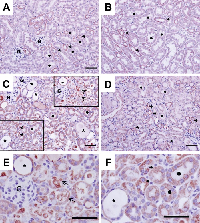
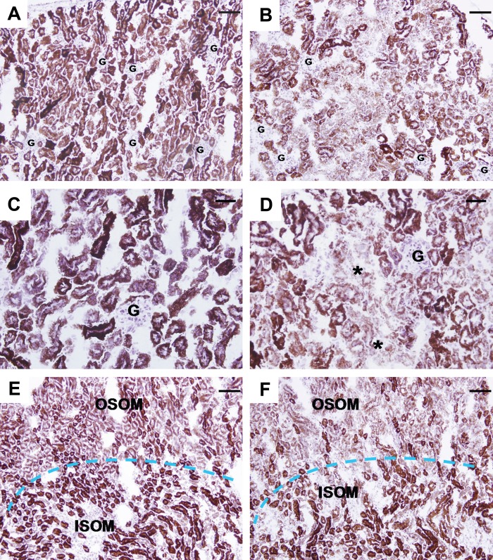
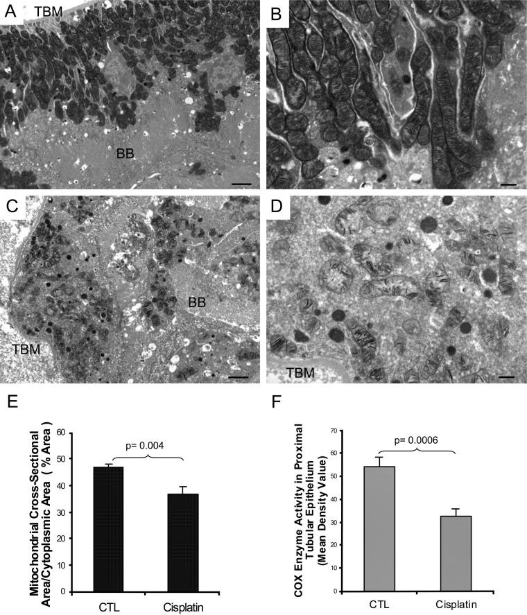
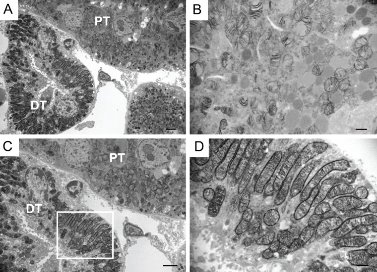
Cisplatin is a widely used antineoplastic agent. However, its major limitation is dose-dependent nephrotoxicity whose precise mechanism is poorly understood. Recent studies have suggested that mitochondrial dysfunction in tubular epithelium contributes to cisplatin-induced nephrotoxicity. Here the authors extend those findings by describing the role of an important electron transport chain enzyme, cytochrome c oxidase (COX). Immunohistochemistry for COX 1 protein demonstrated that, in response to cisplatin, expression was mostly maintained in focally damaged tubular epithelium. In contrast, COX enzyme activity in proximal tubules (by light microscopy) was decreased. Ultrastructural analysis of the cortex and outer stripe of the outer medulla showed decreased mitochondrial mass, disruption of cristae, and extensive mitochondrial swelling in proximal tubular epithelium. Functional electron microscopy showed that COX enzyme activity was decreased in the remaining mitochondria in the proximal tubules but maintained in distal tubules. In summary, cisplatin-induced nephrotoxicity is associated with structural and functional damage to the mitochondria. More broadly, using functional electron microscopy to measure mitochondrial enzyme activity may generate mechanistic insights across a spectrum of renal disorders.


Conflict of interest statement

Declaration of Conflicting Interests: The authors declared no potential conflicts of interest with respect to the research, authorship, and/or publication of this article.

Figures

Figure 1.
Figure 1.
COX 1 immunohisto-chemistry in mouse kidney 72 hr after cisplatin treatment. Panels A and B: control sham-treated mouse kidney. Extensive immunostaining (red stain) is observed in both proximal and distal tubular epithelium; staining is mainly in the basolateral surface of the epithelia. In the cortex (Panel A), distal tubules (arrowheads) are more extensively stained for COX 1 than are proximal epithelium (denoted by ●). In the medulla (Panel B), distal tubules (arrowheads) also exhibit more intense staining than S3 segments of proximal tubules. Panels C, D, E, and F: Cisplatin induced profound histopathological renal injury, desquamation of epithelial cells evidenced by protein cast in the tubular lumen (arrows), and dilation of the tubules (denoted by *). COX 1 expression was mildly decreased in damaged tubular epithelial cells (Panel E), but immunostaining is maintained at a relatively normal distribution in spared epithelium (Panel F). Bars: A, B, C, D: 50 µm.
Figure 2.
Figure 2.
COX enzyme histo-chemistry in mouse kidney 72 hr after cisplatin treatment. Panels A and C (cortex) and E (medulla): Staining (brown) for cytochrome c oxidase enzyme activity on sections of snap-frozen kidney from sham-treated mice. The cortex (A) and inner stripe of the outer medulla (ISOM) have the most intense staining, followed by the outer stripe of the outer medulla (OSOM), with the weakest activity in the inner medulla. Panels B and D (cortex) and F (medulla): Enzyme activity in the cortex and OSOM is greatly decreased 72 hr after cisplatin treatment but unchanged in the ISOM. G: glomerulus. Bars: A, B, E, and F: 80 µm; C and D: 40 µm.
Figure 3.
Figure 3.
Ultrastructural analysis of mitochondrial cytochrome c oxidase enzyme activity in mouse kidney 72 hr after cisplatin treatment. Functional electron microscopy of the renal cortex in sham-treated control (Panels A and B) shows intact proximal tubular epithelium with preserved mitochondria and intense COX activity in every mitochondria of the cell. In cisplatin-treated kidney (Panels C and D), there is extensive acute injury in the proximal tubule. Disorganized mass of mitochondria, with disrupted cristae and greatly reduced COX activity, is seen. TBM: tubular basement membrane, BB: brush border. Bars in Panels A and C: 2 µm; bars in Panels B and D: 500 nm. Panel E: Morphometric analysis of the mitochondrial volume per proximal tubular cytoplasmic area. Percentage mitochondrial area in tubular epithelium is decreased 72 hr after cisplatin treatment compared to control sham-injected mouse kidney. Data are presented as mean ± SEM. * p<0.05, significantly different from sham control. Panel F: Morphometric analysis of COX enzyme activity in mitochondria of the proximal tubular epithelium. COX enzymatic product, measured by mean density value, declined after cisplatin administration compared to control sham-injected mouse kidney. Data are presented as mean ± SEM. ** p<0.005, significantly different from sham control.
Figure 4.
Figure 4.
Comparison of different tubular segments in mouse kidneys 72 hr after cisplatin treatment. Ultrastructural analysis of COX enzyme activity in proximal vs distal tubules of a cisplatin-treated mouse (Panels A and C). In proximal tubules, activity is clearly decreased (Panel B), while in distal tubules, COX enzyme activity is maintained (Panel D). DT: distal tubule, PT: proximal tubule. Bars in Panels A and C: 2 µm; bars in Panels B and D: 500 nm.

References

    1. Aggarwal SK. 1993. A histochemical approach to the mechanism of action of cisplatin and its analogues. J Histochem Cytochem. 7:1053–1073 - PubMed
    1. Anderson WA, Bara G, Seligman AM. 1975. The ultrastructural localization of cytochrome oxidase via cytochrome. J Histochem Cytochem. 1:13–20 - PubMed
    1. Bertoni-Freddari C, Fattoretti P, Casoli T, Di Stefano G, Solazzi M, Gracciotti N, Pompei P. 2001. Mapping of mitochondrial metabolic competence by cytochrome oxidase and succinic dehydrogenase cytochemistry. J Histochem Cytochem. 9:1191–1192 - PubMed
    1. Brady HR, Kone BC, Stromski ME, Zeidel ML, Giebisch G, Gullans SR. 1990. Mitochondrial injury: an early event in cisplatin toxicity to renal proximal tubules. Am J Physiol. 258(5)(Pt 2):F1181–F1187 - PubMed
    1. Brooks C, Wei Q, Cho SG, Dong Z. 2009. Regulation of mitochondrial dynamics in acute kidney injury in cell culture and rodent models. J Clin Invest. 119:1275–1285 - PMC - PubMed

Publication types

MeSH terms