. 2012 Jun 1;287(23):19029-39.
doi: 10.1074/jbc.M112.363390. Epub 2012 Apr 16.

Vacuolar H+-ATPase (V-ATPase) promotes vacuolar membrane permeabilization and nonapoptotic death in stressed yeast

Affiliations

Vacuolar H+-ATPase (V-ATPase) promotes vacuolar membrane permeabilization and nonapoptotic death in stressed yeast

Hyemin Kim et al. J Biol Chem. .

Abstract

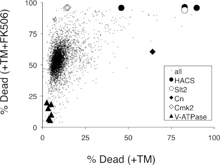
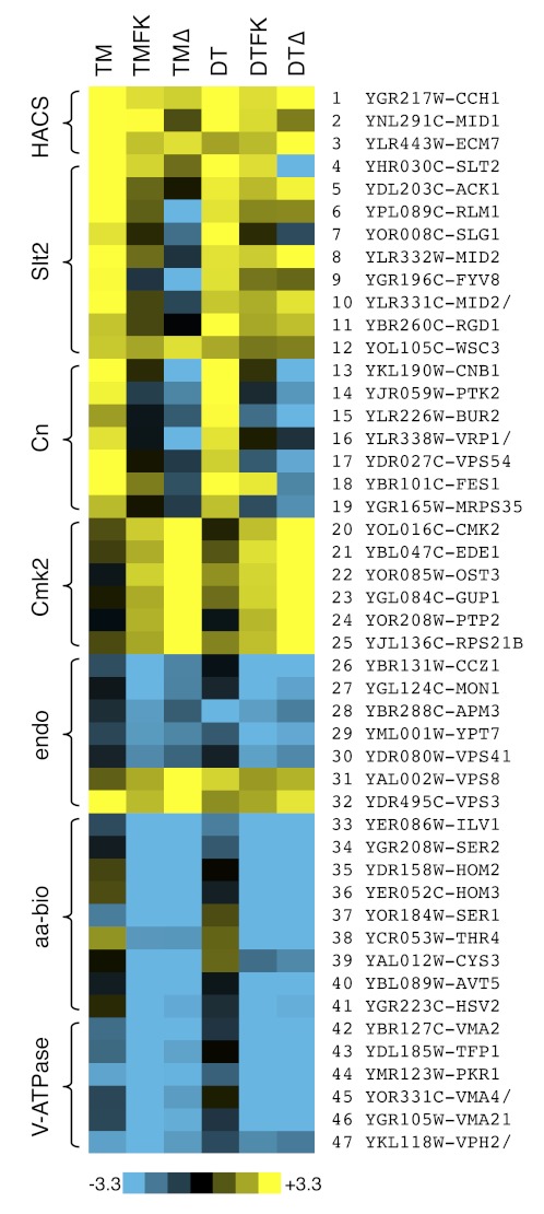
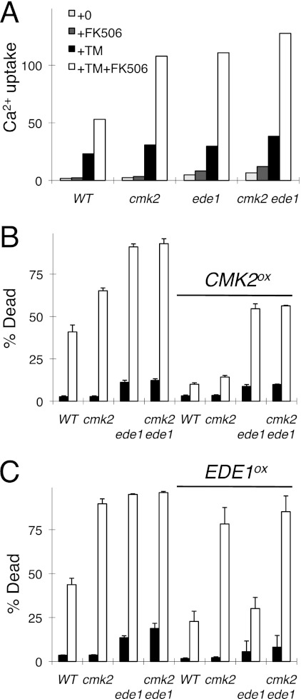
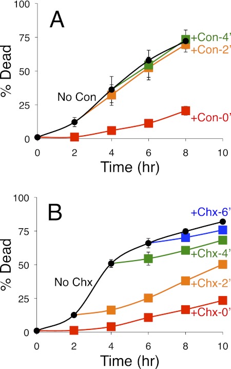
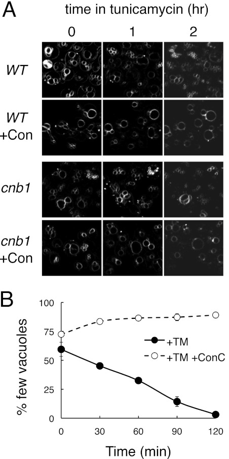
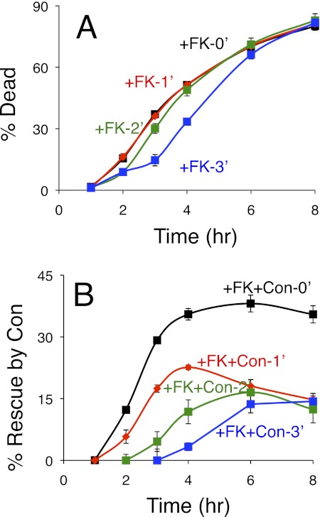
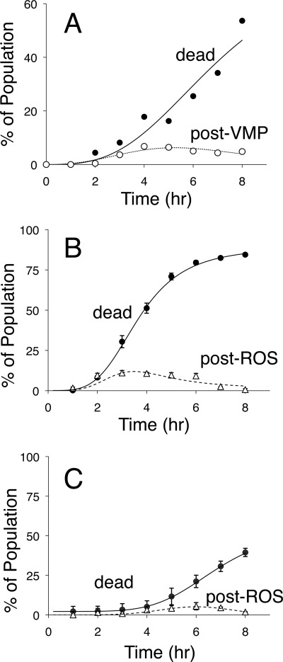
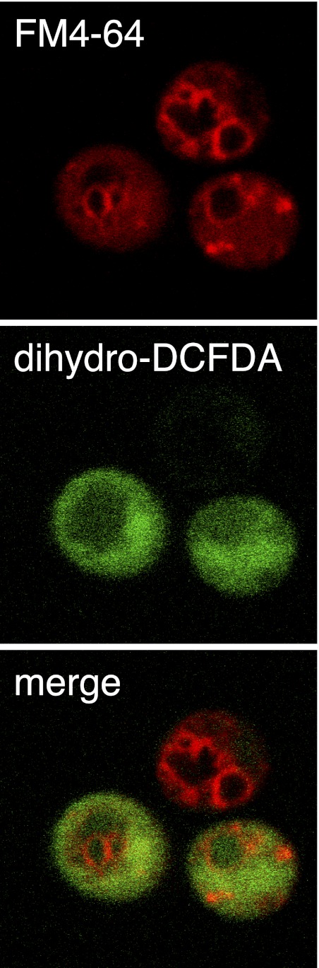
Stress in the endoplasmic reticulum caused by tunicamycin, dithiothreitol, and azole-class antifungal drugs can induce nonapoptotic cell death in yeasts that can be blocked by the action of calcineurin (Cn), a Ca(2+)-dependent serine/threonine protein phosphatase. To identify additional factors that regulate nonapoptotic cell death in yeast, a collection of gene knock-out mutants was screened for mutants exhibiting altered survival rates. The screen revealed an endocytic protein (Ede1) that can function upstream of Ca(2+)/calmodulin-dependent protein kinase 2 (Cmk2) to suppress cell death in parallel to Cn. The screen also revealed the vacuolar H(+)-ATPase (V-ATPase), which acidifies the lysosome-like vacuole. The V-ATPase performed its death-promoting functions very soon after imposition of the stress and was not required for later stages of the cell death program. Cn did not inhibit V-ATPase activities but did block vacuole membrane permeabilization (VMP), which occurred at late stages of the cell death program. All of the other nondying mutants identified in the screens blocked steps before VMP. These findings suggest that VMP is the lethal event in dying yeast cells and that fungi may employ a mechanism of cell death similar to the necrosis-like cell death of degenerating neurons.


Figures

FIGURE 1.
FIGURE 1.
Genome-wide screen for mutants with altered rates of cell death. Individual mutants from the yeast gene knock-out collection were cultured in the presence of tunicamycin (+TM) and tunicamycin plus FK506 (+TM+FK506), stained with PI, analyzed by flow cytometry, and plotted as a single point in the chart. Each point represents the percentage of dead cells in the culture containing tunicamycin versus that in the culture containing tunicamycin plus FK506. Selected mutants have been highlighted with large symbols.
FIGURE 2.
FIGURE 2.
Heat maps of cell death for selected mutants. Raw data from Fig. 1 and a secondary screen involving dithiothreitol (DT) instead of tunicamycin (TM) were converted to Z-scores as described under “Experimental Procedures,” and mutants with patterns of cell death similar to or opposite from those of HACS-, Cn-, and Cmk2-deficient cells are illustrated. The scale bar illustrates the number of standard deviations below (blue) or above (yellow) the population averages. TMΔ and DTΔ represent Z-scores of the FK506 responsiveness of each mutant strain.
FIGURE 3.
FIGURE 3.
Ede1 stimulates Cmk2 functions. A–C, mutants lacking Ede1, Cmk2, or both proteins were compared with wild-type yeast in assays of 45Ca2+ uptake (A) and cell death (B and C) in response to tunicamycin (TM) and FK506. The strains bearing control plasmids (left half of B and C) or Cmk2 and Ede1 overexpression plasmids (right half of B and C) were analyzed in parallel. Error bars indicate ±S.D.
FIGURE 4.
FIGURE 4.
Inhibition of cell death by inhibitors of V-ATPase and translation. A and B, cultures of the Cn-deficient cnb1 mutants were exposed to tunicamycin at 0 h with additions of either the V-ATPase inhibitor concanamycin (Con) (A) or the translation inhibitor cycloheximide (Chx) (B) at 0 h (red), 2 h (orange), 4 h (green), 6 h (blue), or never (black). The frequencies of cell death in triplicate cultures were measured at the indicated times by staining with PI and flow cytometry. Error bars indicate ±S.D.
FIGURE 5.
FIGURE 5.
V-ATPase-dependent vacuole fragmentation in response to tunicamycin. A, wild-type and Cn-deficient cnb1 mutants were stained with the fluorescent dye FM4-64 to reveal vacuole membranes and imaged in a microfluidic device at various times of exposure to tunicamycin or tunicamycin plus concanamycin (+Con) as indicated. B, wild-type cells prestained with FM4-64 were imaged by fluorescence microscopy at various time points after exposure to tunicamycin (+TM) or tunicamycin plus FK506 (+TM+Con) and counted manually as having few vacuoles (two or fewer) or several vacuoles (three or more). The averages of three parallel experiments (±S.D.) were charted.
FIGURE 6.
FIGURE 6.
Cn delays V-ATPase-dependent cell death. Cultures of wild-type yeast cells were exposed to tunicamycin at 0 h with additions of FK506 (FK) or FK506 plus concanamycin (FK+Con) at 0 h (black), 1 h (red), 2 h (green), or 3 h (blue). A and B, the frequencies of cell death in triplicate cultures were measured at the indicated times by staining with PI and flow cytometry and plotted directly as percentage of dead (A) or plotted after determining the percentage of the population that was rescued by the addition of concanamycin (B), which was calculated by subtraction of the values obtained in the FK506 plus concanamycin cultures from those shown in A. Error bars indicate ±S.D.
FIGURE 7.
FIGURE 7.
Vacuole membrane permeabilization and ROS accumulation in dying populations of yeast cells. A, the Cn-deficient cnb1 mutant was exposed to tunicamycin, incubated at room temperature, stained at the indicated times with either PI or carboxy-DCFDA, and immediately observed by epifluorescence microscopy. The frequency of dead cells in the population that stained with PI was plotted (black circles), and the frequency of cells in the population that failed to stain with PI and exhibited cytoplasmic staining with carboxy-DCFDA was plotted (white circles). B and C, wild-type (B) and V-ATPase-deficient vma1 mutant cells (C) were exposed to tunicamycin plus FK506, stained with PI at the indicated times, counted by flow cytometry, and plotted as the average (±S.D.) from the three replicate experiments (black circles). The same cultures were also sampled, stained with dihydro-DCFDA at the indicated times, counted manually in the fluorescence microscope, and plotted (white triangles). The PI staining data were fit by nonlinear regression to the standard sigmoid equation (solid lines), and the other staining data were similarly to fit to the difference between a standard sigmoid equation and the solid lines (dashed lines).
FIGURE 8.
FIGURE 8.
Nonvacuolar localization of ROS. Wild-type yeast cells were stained with FM4-64 and subsequently exposed to tunicamycin plus FK506. After 4 h of incubation, cells were stained with dihydro-DCFDA and imaged by confocal microscopy for vacuole membrane architecture (FM4-64) and ROS accumulation (dihydro-DCFDA). The image shows three live cells, two of which are representative of cells that stain positive for ROS. Fluorescence products of dihydro-DCFDA were almost always excluded from the region of the cell containing vacuoles.

References

    1. Kroemer G., Galluzzi L., Vandenabeele P., Abrams J., Alnemri E. S., Baehrecke E. H., Blagosklonny M. V., El-Deiry W. S., Golstein P., Green D. R., Hengartner M., Knight R. A., Kumar S., Lipton S. A., Malorni W., Nuñez G., Peter M. E., Tschopp J., Yuan J., Piacentini M., Zhivotovsky B., Melino G. (2009) Classification of cell death: recommendations of the Nomenclature Committee on Cell Death 2009. Cell Death Differ. 16, 3–11 - PMC - PubMed
    1. Golstein P., Kroemer G. (2007) Cell death by necrosis: toward a molecular definition. Trends Biochem. Sci. 32, 37–43 - PubMed
    1. Driscoll M., Gerstbrein B. (2003) Dying for a cause: invertebrate genetics takes on human neurodegeneration. Nat. Rev. Genet. 4, 181–194 - PubMed
    1. Syntichaki P., Xu K., Driscoll M., Tavernarakis N. (2002) Specific aspartyl and calpain proteases are required for neurodegeneration in C. elegans. Nature 419, 939–944 - PubMed
    1. Syntichaki P., Samara C., Tavernarakis N. (2005) The vacuolar H+-ATPase mediates intracellular acidification required for neurodegeneration in C. elegans. Curr. Biol. 15, 1249–1254 - PubMed

Publication types

MeSH terms

Substances

LinkOut - more resources