

Case Reports
doi: 10.3350/kjhep.2012.18.1.101.
Epub 2012 Mar 22.
MR imaging of hepatic lymphangioma
Affiliations
- PMID: 22511911
- PMCID: PMC3326998
- DOI: 10.3350/kjhep.2012.18.1.101
Item in Clipboard
Case Reports
MR imaging of hepatic lymphangioma
Korean J Hepatol.
2012 Mar.
No abstract available
Keywords: CT; Liver; Lymphangioma; MRI; Ultrasound.
Figures
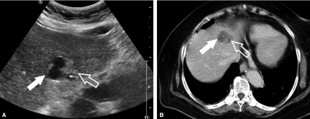
A mixed echoic hepatic nodule in the segment IV is seen on ultrasonography. The right side of the lesion is anechoic (solid arrow), and the left is hyperechoic (open arrow) (A). On the post enhanced CT scan shows the right side of the lesions is a relatively well-circumscribed low attenuated portion (solid arrow), but the other side is enhanced heterogeneously (open arrow). This feature is matched with ultrasonographic findings (B).

MR scan shows that its features on the 3D-spoiled gradient echo T1-weighted MR scan after gadolinium contrast injection are very similar to the that of CT scan (A), and fat-suppressed T2-weighted MR scan shows a bright high signal intensity in the whole mass (solid and open arrows), differentiated from the US and CT features. This finding suggests that this hepatic mass may be composed of a fluid-filled structure, cyst (B). Coronal T2-weighted MR scan demonstrates the aggregated cystic component on the left side of the mass well (open arrow) (C).
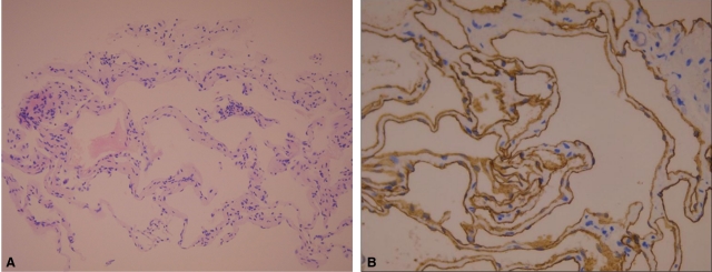
Pathologically, the sponge-like lesion is composed of microscopic cysts lined by attenuated endothelium, resembling that in normal lymphatics (H-E stain, ×20) (A). Immunohistochemical staining with CD34 antibody, reveals the lining endothelium is well demonstrated on the microscopy (CD34 stain, ×400) (B).
References
-
- Koh CC, Sheu JC. Hepatic lymphangioma--a case report. Pediatr Surg Int. 2000;16:515–516. - PubMed
-
- Chan SC, Huang SF, Lee WC, Wan YL. Solitary hepatic lymphangioma--a case report. Int J Clin Pract Suppl. 2005;(147):100–102. - PubMed
-
- Lugo-Olivieri CH, Taylor GA. CT differentiation of large abdominal lymphangioma from ascites. Pediatr Radiol. 1993;23:129–130. - PubMed
-
- Ko SF, Ng SH, Shieh CS, Lin JW, Huang CC, Lee TY. Mesenteric cystic lymphangioma with myxoid degeneration: unusual CT and MR manifestations. Pediatr Radiol. 1995;25:525–527. - PubMed
Publication types
MeSH terms
Substances
LinkOut - more resources
Full Text Sources