. 2012 May 1;28(17):7063-70.
doi: 10.1021/la300566x. Epub 2012 Apr 18.

DNA electrochemistry with tethered methylene blue

Affiliations

DNA electrochemistry with tethered methylene blue

Catrina G Pheeney et al. Langmuir. .

Abstract

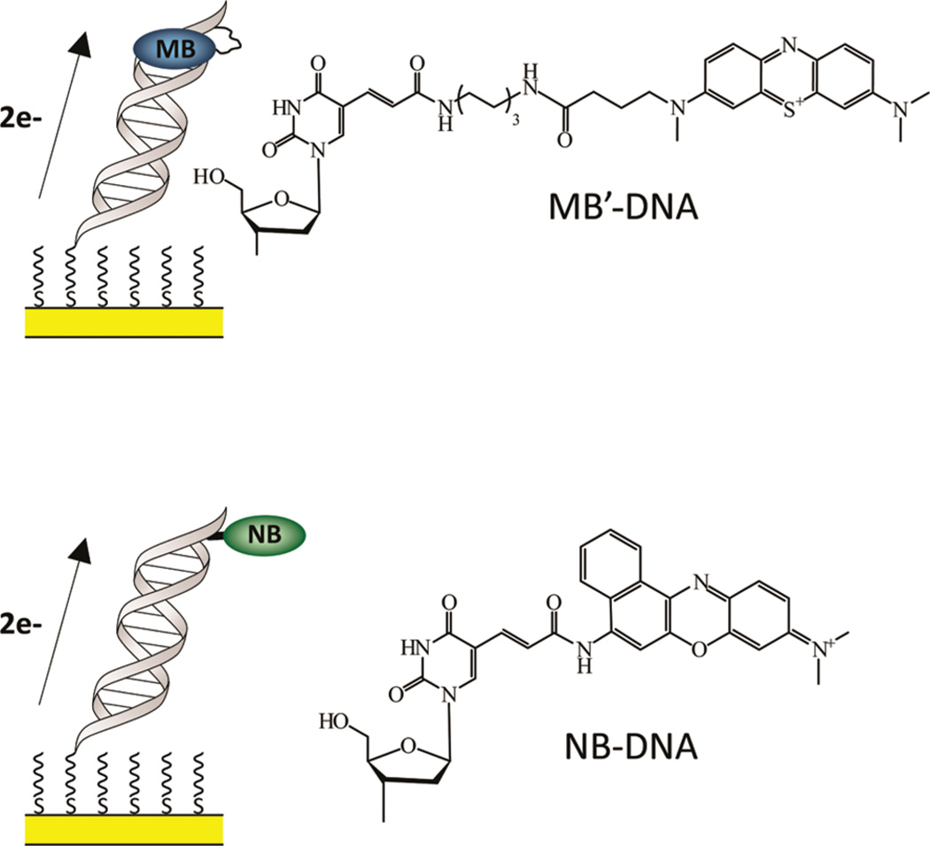
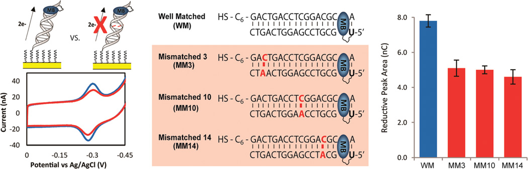
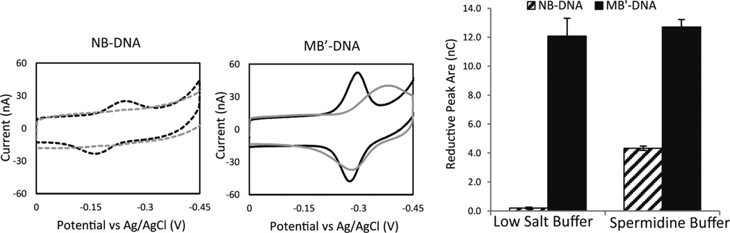
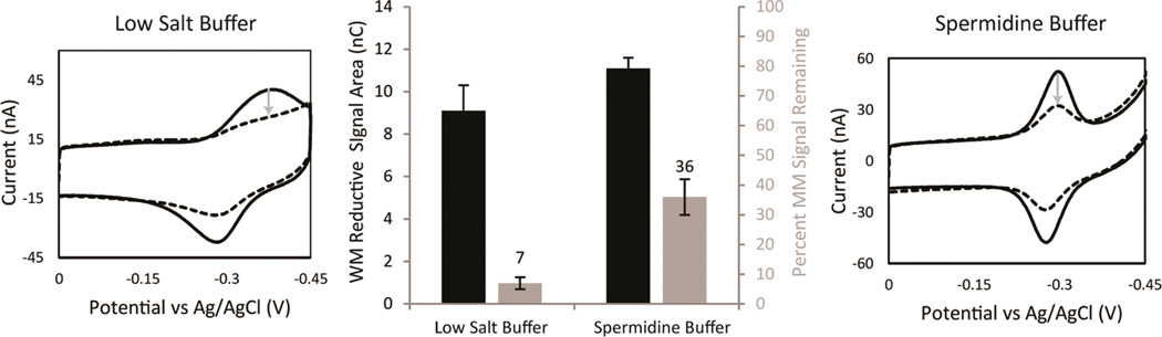
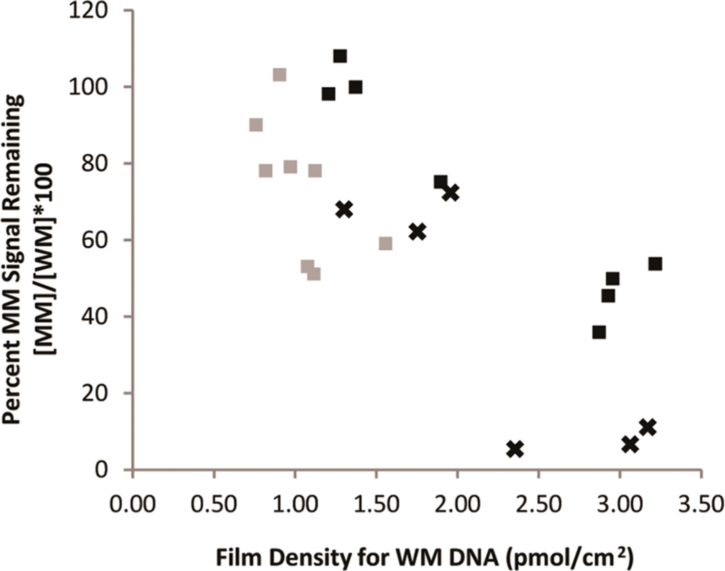
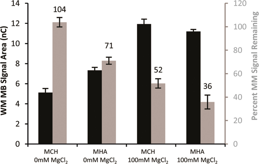
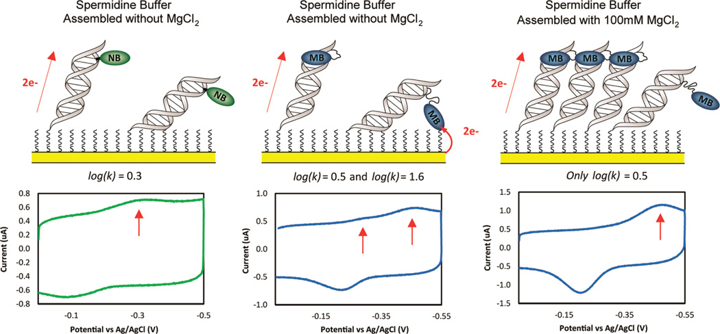
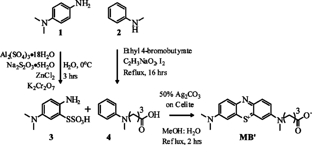
Methylene blue (MB'), covalently attached to DNA through a flexible C(12) alkyl linker, provides a sensitive redox reporter in DNA electrochemistry measurements. Tethered, intercalated MB' is reduced through DNA-mediated charge transport; the incorporation of a single base mismatch at position 3, 10, or 14 of a 17-mer causes an attenuation of the signal to 62 ± 3% of the well-matched DNA, irrespective of position in the duplex. The redox signal intensity for MB'-DNA is found to be least 3-fold larger than that of Nile blue (NB)-DNA, indicating that MB' is even more strongly coupled to the π-stack. The signal attenuation due to an intervening mismatch does, however, depend on DNA film density and the backfilling agent used to passivate the surface. These results highlight two mechanisms for reduction of MB' on the DNA-modified electrode: reduction mediated by the DNA base pair stack and direct surface reduction of MB' at the electrode. These two mechanisms are distinguished by their rates of electron transfer that differ by 20-fold. The extent of direct reduction at the surface can be controlled by assembly and buffer conditions.


Figures

Figure 1
Figure 1
Left: schematic illustration of MB′–DNA (top) and NB–DNA (bottom) monolayers bound to an electrode. The reporter is appended to the distal base of the duplex. The intended path for electrochemical reduction is indicated. Right: the chemical structure of both MB′ and NB covalently tethered to a modified uracil.
Figure 2
Figure 2
Electrochemistry of MB′–DNA with a single intervening CA mismatch. Left: cyclic voltammetry was acquired at 100 mV/s with either a well-matched sequence (blue) or a sequence containing a single mismatch (red). All four sequences (middle) of MB′–DNA were assembled on the same multiplexed chip and assembled without MgCl2, passivated with MHA, and scanned in spermidine buffer. Right: the area of the reductive signal was used to quantify the signals observed, and the errors denoted are determined from the variation across four different electrodes.
Figure 3
Figure 3
Comparison of the signal from NB–DNA (dotted) and MB′–DNA (solid). The electrodes were assembled with 100 mM MgCl2 and passivated with MCH. CVs were acquired at 100 mV/s, and all electrodes were scanned in both a low salt buffer (5.0 mM phosphate, 50 mM NaCl, and pH 7) (gray) and a spermidine buffer (5.0 mM phosphate, 50 mM NaCl, 4 mM MgCl2, 4 mM spermidine, 50 µM EDTA, 10% glycerol, and pH 7) (black). The CVs shows the overall signal changes in response to the running buffer. The reductive peak areas were quantified to show that the amount of MB′–DNA being reduced is relatively unchanged regardless of the buffer. The errors denoted are determined from the variation across four different electrodes.
Figure 4
Figure 4
Optimization of mismatch discrimination depending on the running buffer. Both WM MB′–DNA (solid) and MM10 MB′–DNA (dotted) modified electrodes were assembled with 100 mM MgCl2, passivated with MHA, and scanned in both a low salt buffer (5.0 mM phosphate, 50 mM NaCl, and pH 7) (right) and a spermidine buffer (5.0 mM phosphate, 50 mM NaCl, 4 mM MgCl2, 4 mM spermidine, 50 µM EDTA, 10% glycerol, and pH 7) (left). The areas of the reductive peaks were used to quantify the reductive signal size (black) and determine the percent signal remaining ([MM]/[WM] × 100) (gray) from the incorporation of a single mismatch. The gray arrows denote the decrease in signal area with the optimal percent signal attenuation being in low salt buffer. The errors denoted were determined by the standard deviation from four electrodes averages, for each sequence of DNA, across three chips.
Figure 5
Figure 5
Dependence of mismatch signal attenuation on film density for both MB′–DNA (black) and NB–DNA (gray). Percent MB′–DNA signal remaining due to the incorporation of a CA mismatch was determined in both low salt buffer (×) and spermidine buffer (squares).
Figure 6
Figure 6
Dependence on assembly conditions of the percent mismatch signal remaining for MB′–DNA. Well-matched signal area (black) and the percent signal remaining (gray) were from 12 electrodes averaged across three chips. The backfilling agent (1 mM MCH or MHA) and concentration of MgCl2 are indicated. Scans were acquired in spermidine buffer.
Figure 7
Figure 7
Top: schematic illustration of reporter reduction mechanisms. Bottom: CV at 5 V/s (bottom) for NB–DNA (left) and MB′–DNA (center) assembled without MgCl2 and MB′–DNA assembled with 100 mM MgCl2 (right). Electrodes were backfilled with mercaptohexanol and scanned in spermidine buffer. The red arrows indicate the peaks quantified in order to determine the rate of the various processes for reporter reduction.
Scheme 1
Scheme 1
Synthetic Strategy for the Preparation of N-Carboxypropyl Methylene Blue (MB′)

References

    1. Murphy CJ, Arkin MR, Jenkins Y, Ghatlia ND, Bossmann SH, Turro NJ, Barton JK. Long Range Photoinduced Electron Transfer through a DNA Helix. Science. 1993;262:1025–1029. - PubMed
    1. Holmlin ER, Dandliker PJ, Barton JK. Charge Transfer Through the DNA Base Stack. Angew. Chem., Int. Ed. 1997;36:2714–2730.
    1. Genereux JC, Boal AK, Barton JK. DNA-Mediated Charge Transport in Redox Sensing and Signaling. J. Am. Chem. Soc. 2010;132:891–905. - PMC - PubMed
    1. Schuster GB, editor. Topics in Current Chemistry. Vol. 237. Berlin: Springer-Verlag; 2004. Long Range Charge Transfer in DNA II; p. 103.
    1. Wagenknecht HA, editor. Charge Transfer in DNA: From Mechanism to Application. Weiheim: Wiley-VCH Verlag GmbH & Co KGaA; 2005.

Publication types