Review
. 2013 Jan;222(1):32-40.
doi: 10.1111/j.1469-7580.2012.01506.x. Epub 2012 Apr 18.

The origin and evolution of the ectodermal placodes

Affiliations
Review

The origin and evolution of the ectodermal placodes

Anthony Graham et al. J Anat. 2013 Jan.

Abstract

Many of the features that distinguish the vertebrates from other chordates are found in the head. Prominent amongst these differences are the paired sense organs and associated cranial ganglia. Significantly, these structures are derived developmentally from the ectodermal placodes. It has therefore been proposed that the emergence of the ectodermal placodes was concomitant with and central to the evolution of the vertebrates. More recent studies, however, indicate forerunners of the ectodermal placodes can be readily identified outside the vertebrates, particularly in urochordates. Thus the evolutionary history of the ectodermal placodes is deeper and more complex than was previously appreciated with the full repertoire of vertebrate ectodermal placodes, and their derivatives, being assembled over a protracted period rather than arising collectively with the vertebrates.


Figures

Fig. 1
Fig. 1
A schematic showing the location of the ectodermal placodes in the vertebrate head. The olfactory placode (olf) coloured red is found at the tip of the forebrain, while the lens (L) placode shaded orange forms caudally as part of the eye. The adenohypophyseal (Ad) placodes, in yellow, can be seen to lie ventrally to the forebrain. The trigeminal placodes form alongside the anterior hindbrain at the levels of rhombomeres 1 and 2. The more anterior ophthalmic (To) placode is coloured dark blue and the maxillomandibular (Tmm) is shaded in a lighter blue. The large otic (Ot) placode forms opposite the central domain of the hindbrain and is in purple. The lateral line (LL) placodes form both anteriorly and posteriorly of the otic vesicle. The epibranchial placodes – geniculate (Eg), petrosal (Ep) and nodose (En) – form as part of the pharyngeal series. They are shaded green. Fore – forebrain, mid – midbrain, r1 – rhombomere 1, r2 – rhombomere 2, r3 – rhombomere 3, r4 – rhombomere 4, pa1 – pharyngeal arch1, pa2, pharyngeal arch 2, pa3 – pharyngeal arch 3, pa4 – pharyngeal arch 4, pa5 – pharyngeal arch s-somite.
Fig. 2
Fig. 2
Phylogenetic tree of the major taxonomic groups in the Deuterostomia. The echinoderms and hemichordates form a group known as the Ambulacraria. Within the chordates, the cephalochordates are the earliest diverging lineages with the urochordates most closely related to the vertebrates.
Fig. 3
Fig. 3
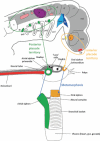
Relationships between vertebrate placodes and putative homologues in a urochordate such as Ciona intestinalis. The top panel is replicated from Fig. 1. In the middle is a schematic diagram of a urochordate tadpole larva with anterior to the right. The notochord is in red and the two siphon primordia are colour coded in green and orange, with putative relationships to subsets of vertebrate placodes shown. At the bottom is an adult urochordate, with blue arrows and colour coding showing what happens to the siphon primordia after metamorphosis.
Fig. 4
Fig. 4
Phylogenetic tree of the Deuterostomia, as shown in Fig. 1. Onto this are marked inferred ancestral placode characters. At the base of the tree, the development of isolated cells from the ectoderm is likely to be very ancient. The anterior pituitary territory is also old; we show two possible placements as it is clearly a chordate wide character (black arrow) but may also predate deuterostome radiation (grey arrow). We suggest paired anterior and posterior thickenings evolved before the separation of the vertebrate and urochordate lineages, with homologies as shown in Fig. 3, with further elaboration of these domains and origin of lens and trigeminal placodes within the vertebrate lineage.

References

    1. Baatrup E. On the structure of the corpuscles of de Quatrefages (Branchiostoma lanceolatum (P)) Acta Zool. 1982;63:39–44.
    1. Bailey AP, Bhattacharyya S, Bronner-Fraser M, et al. Lens specification is the ground state of all sensory placodes, from which FGF promotes olfactory identity. Dev Cell. 2006;11:505–517. - PubMed
    1. Baker CV, Bronner-Fraser M. Vertebrate cranial placodes. I. Embryonic induction. Dev Biol. 2001;232:1–61. - PubMed
    1. Barraud P, Seferiadis AA, Tyson LD, et al. Neural crest origin of olfactory ensheathing glia. Proc Natl Acad Sci U S A. 2010;107:21040–21045. - PMC - PubMed
    1. Bassham S, Postlethwait JH. The evolutionary history of placodes: a molecular genetic investigation of the larvacean urochordate Oikopleura dioica. Development. 2005;132:4259–4272. - PubMed

LinkOut - more resources