

Case Reports
doi: 10.1056/NEJMoa1110813.
Integrin α3 mutations with kidney, lung, and skin disease
Affiliations
- PMID: 22512483
- PMCID: PMC3341404
- DOI: 10.1056/NEJMoa1110813
Item in Clipboard
Case Reports
Integrin α3 mutations with kidney, lung, and skin disease
N Engl J Med.
.
Abstract
Integrin α(3) is a transmembrane integrin receptor subunit that mediates signals between the cells and their microenvironment. We identified three patients with homozygous mutations in the integrin α(3) gene that were associated with disrupted basement-membrane structures and compromised barrier functions in kidney, lung, and skin. The patients had a multiorgan disorder that included congenital nephrotic syndrome, interstitial lung disease, and epidermolysis bullosa. The renal and respiratory features predominated, and the lung involvement accounted for the lethal course of the disease. Although skin fragility was mild, it provided clues to the diagnosis.
Figures
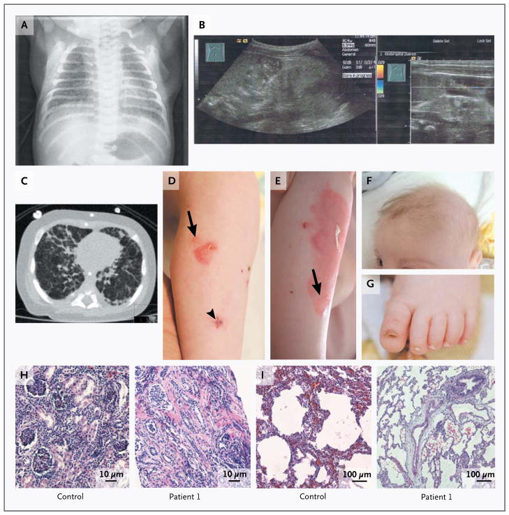
Panel A shows a chest radiograph obtained on the first day of life. Panel B shows the results of neonatal ultrasonography, revealing bilateral, orthotopic small kidneys with hyperechogenic parenchyma, without corticomedullary differentiation, suggesting renal dysplasia. Panel C shows a computed tomographic scan of the chest obtained at the age of 4 months, revealing diffuse distortion of the pulmonary architecture, coarsened interstitial changes in the lung with thickened interlobular and intralobular septa in all segments; other images showed consolidation in the right posterior upper lobe. Panels D and E show progressive skin blisters and erosions on the leg (arrows), with an interval of 30 days between the two photographs. The arrowhead indicates the biopsy site. Panel F shows very fine, sparse scalp and eyebrow hair, and Panel G shows a dystrophic toenail. Panel H shows kidney-biopsy samples obtained from a control subject and Patient 1, revealing atrophic glomeruli, single collapsed loops, segmental sclerosis, occasionally fibrous obliteration of Bowman’s capsule space, and focal tubular atrophy in the patient. Diffuse interstitial fibrosis is associated with lymphocytic infiltrates. Panel I shows lung-biopsy specimens obtained from a control subject and Patient 1, revealing well-expanded lung parenchyma with irregular air-space expansion, areas of alveolar duct distention, and zones of mild-to-moderate peripheral air-space distention and simplification, with intact respiratory epithelium, in the patient (hematoxylin and eosin staining in Panels H and I).
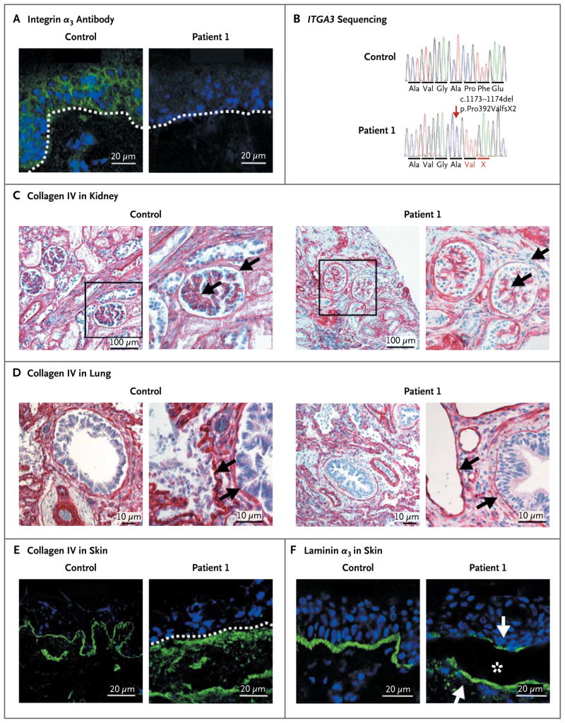
In Panel A, immunofluorescence (green) staining of a skin specimen with the monoclonal integrin α3 antibody P1B5 shows loss of expression; a skin specimen from a control subject is shown for comparison. The position of the epidermal basement membrane is indicated by the white dashed line; nuclei are blue. In Panel B, chromatograms show the partial sequences of ITGA3. The homozygous mutation c.1173_1174del (arrow) leads to frameshift and formation of a premature termination codon, p.Pro392ValfsX2. Immunohistochemical and immunofluorescence staining of collagen IV is shown in biopsy specimens of kidney (Panel C), lung (Panel D), and skin (Panel E) obtained from Patient 1 and age-matched controls. In Panel C, the marked areas in the images on the left side are shown at higher magnification on the right side; the arrows indicate the glomerular basement membrane and Bowman’s capsule basement membrane. In addition, there is a loss of lateral cell junctions in the patient. In Panel D, arrows point to the basement membranes, which are very thin in the specimen from the patient. Panel E shows irregularly distributed collagen IV below the basement membrane (dashed white line) in the patient’s skin. Panel F shows immunofluorescence staining of the laminin α3 chain, a ligand of integrin α3. In the patient’s skin, laminin α3 appears at the blister (asterisk) roof and base (arrows).
References
-
- Danen EH, Sonnenberg A. Integrins in regulation of tissue development and function. J Pathol. 2003;200:471–80. [Corrected and republished, J Pathol 2003;201: 632–41.] - PubMed
-
- Kambham N, Tanji N, Seigle RL, et al. Congenital focal segmental glomerulosclerosis associated with beta4 integrin mutation and epidermolysis bullosa. Am J Kidney Dis. 2000;36:190–6. - PubMed
-
- Margadant C, Charafeddine RA, Sonnenberg A. Unique and redundant functions of integrins in the epidermis. FASEB J. 2010;24:4133–52. - PubMed
Publication types
MeSH terms
Substances
Grants and funding
LinkOut - more resources
Full Text Sources
Other Literature Sources
Medical
Molecular Biology Databases