Review
. 2012 Nov 16:1485:40-53.
doi: 10.1016/j.brainres.2012.03.044. Epub 2012 Mar 27.

Understanding tinnitus: the dorsal cochlear nucleus, organization and plasticity

Affiliations
Review

Understanding tinnitus: the dorsal cochlear nucleus, organization and plasticity

Joan S Baizer et al. Brain Res. .

Abstract

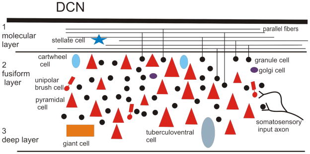
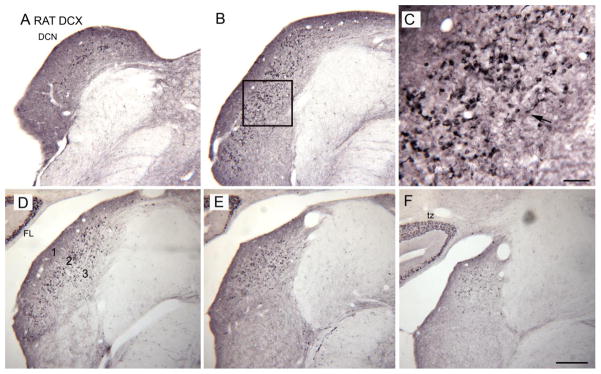
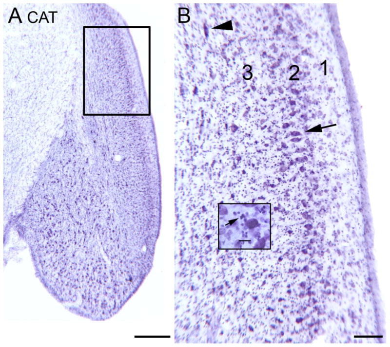
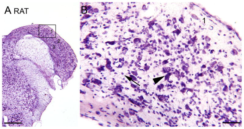
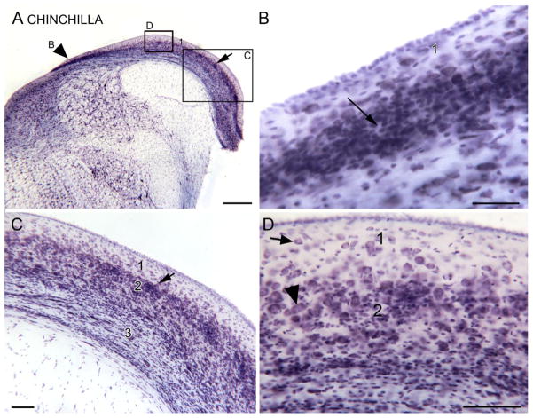
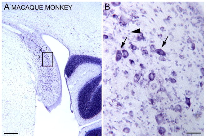
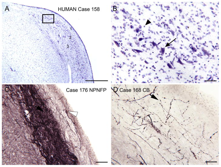
Tinnitus, the perception of a phantom sound, is a common consequence of damage to the auditory periphery. A major goal of tinnitus research is to find the loci of the neural changes that underlie the disorder. Crucial to this endeavor has been the development of an animal behavioral model of tinnitus, so that neural changes can be correlated with behavioral evidence of tinnitus. Three major lines of evidence implicate the dorsal cochlear nucleus (DCN) in tinnitus. First, elevated spontaneous activity in the DCN is correlated with peripheral damage and tinnitus. Second, there are somatosensory inputs to the DCN that can modulate spontaneous activity and might mediate the somatic-auditory interactions seen in tinnitus patients. Third, we have found a subpopulation of DCN neurons in the adult rat that express doublecortin, a plasticity-related protein. The expression of this protein may reflect a role of these neurons in the neural reorganization causing tinnitus. However, there is a problem in extending the findings in the rodent DCN to humans. Classic studies state that the structure of the primate DCN is quite different from that of rodents, with primates lacking granule cells, the recipients of somatosensory input. To address the possibility of major species differences in DCN organization, we compared Nissl-stained sections of the DCN in five different species. In contrast to earlier reports, our data suggest that the organization of the primate DCN is not dramatically different from that of the rodents, and validate the use of animal data in the study of tinnitus. This article is part of a Special Issue entitled: Tinnitus Neuroscience.


Figures

Figure 1
Figure 1
Laminar and cellular organization of the DCN, based on Nissl-stained sections in the rat. Only somata are represented. Stellate, cartwheel, and Golgi cells are inhibitory interneurons. Granule cells are the source of the excitatory parallel fibers in the molecular layer (1). The giant cells, tuberculoventral cells and pyramidal cells are excitatory projection neurons, and the unipolar brush cells are excitatory interneurons.
Figure 2
Figure 2
A, B, D– F. DCX expression in the DCN on five sections about 250 μm apart. A is the most caudal section. C. The DCX-ir profiles are UBCs. The arrow indicates one example in which the soma and brush are both visible in the plane of section.
Figure 3
Figure 3
The DCN in the cat shown on a CV-stained section. A. Low magnification image; note the laminar appearance. The rectangle shows the area in the larger image in B. Scale bar = 500 μm B. Laminar organization of cat DCN. There is much lighter staining in the outer or molecular layer (1). The arrow indicates a group of neurons in the fusiform/pyramidal cell layer (2) with elongated cell bodies oriented perpendicular to the surface. The arrowhead shows a cell in the polymorphic (3) layer with an elongated cell body. Scale bar =100 μm. The inset shows small, darkly labeled profiles, at arrow. Scale bar in inset = 0 μm.
Figure 4
Figure 4
The DCN in the rat, CV staining. A. Low magnification image. The rectangle shows the location of the higher magnification image in B. Scale bar = 500 μm. B. 1, 2, 3, indicate the layers. The arrowhead shows a large cell with fusiform cell body oriented perpendicular to the surface. The arrow shows small neurons.
Figure 5
Figure 5
The DCN in the chinchilla, CV staining. A. Low magnification image of the DCN. The arrowhead on the left indicates a dense band of darkly stained small cells and is an alignment point for the higher magnification image in Fig. 4B. Scale bar = 500 μm. B. Dense band of stained granule cells, examples at arrow. Scale bar = 50 μm.
Figure 6
Figure 6
The DCN in the macaque monkey, CV staining. A. Laminar organization (three layers, 1, 2, and 3 can be distinguished) is visible at low magnification. The rectangle shows the location of the higher magnification image in B. Scale bar=500 μm. B. Image through the fusiform/pyramidal cell layer (2). The arrows indicate the elongated somata of larger neurons oriented at different angles. The arrowhead indicates two small profiles. Scale bar=50 μm.
Figure 7
Figure 7
The DCN in the human. A. CV staining. There is an outer layer (1) with few stained somata, a darker band below it (2) with scattered, stained, large somata, and lighter staining and more scattered profiles deep to that (3). The boxed region is shown at higher magnification in B. Scale bar=1 mm. B. Large stained profiles; somata are elongated but not all oriented the same way. Note the many small stained somata (arrowhead). Scale bar=50 μm. C. Laminar organization as shown by label with an antibody to nonphosphorylated neurofilament protein (NPNFP). There is a darkly stained band made up of somata (example at arrow) and a dense meshwork of stained processes. In layer 1 there one stained neuron and a few stained fibers (example at arrowhead). Scale bar=50 μm. D. Calbindin (CB)-immunoreactivity in fine, beaded fibers (example at arrowhead) that overlap layer 2. Scale bar= 50 μm.

References

    1. Abbott SD, Hughes LF, Bauer CA, Salvi R, Caspary DM. Detection of glutamate decarboxylase isoforms in rat inferior colliculus following acoustic exposure. Neuroscience. 1999;93:1375–81. - PubMed
    1. Adams JC. Neuronal morphology in the human cochlear nucleus. Arch Otolaryngol Head Neck Surg. 1986;112:1253–61. - PubMed
    1. Adjamian P, Sereda M, Hall DA. The mechanisms of tinnitus: perspectives from human functional neuroimaging. Hear Res. 2009;253:15–31. - PubMed
    1. Aitkin LM, Phillips SC. Is the inferior colliculus an obligatory relay in the cat auditory system? Neurosci Lett. 1984;44:259–64. - PubMed
    1. Aitkin LM, Pettigrew JD, Calford MB, Phillips SC, Wise LZ. Representation of stimulus azimuth by low-frequency neurons in inferior colliculus of the cat. J Neurophysiol. 1985;53:43–59. - PubMed

Publication types

Substances