Review
. 2012 Jan-Feb;6(1):1-10.
doi: 10.4161/chan.19126. Epub 2012 Jan 1.

KCNE2 and the K (+) channel: the tail wagging the dog

Affiliations
Review

KCNE2 and the K (+) channel: the tail wagging the dog

Geoffrey W Abbott. Channels (Austin). 2012 Jan-Feb.

Abstract

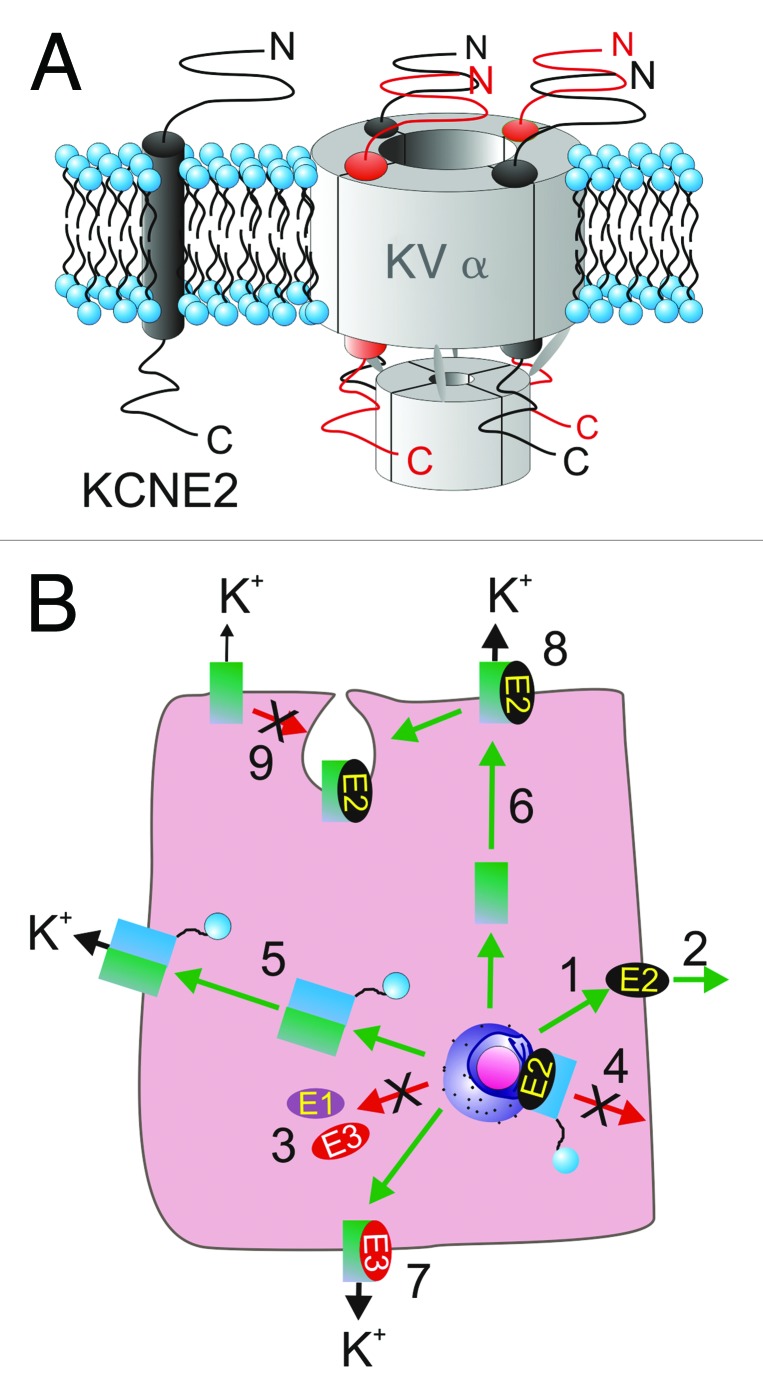
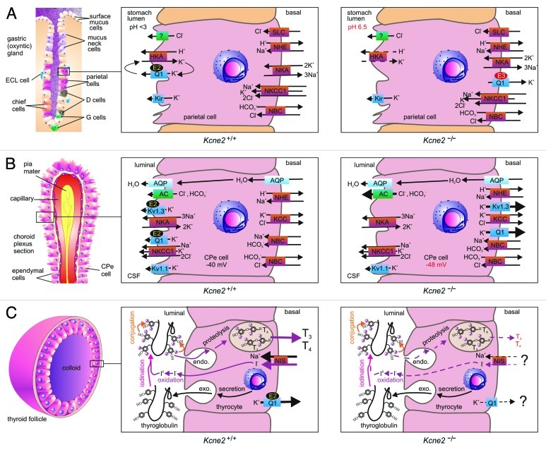
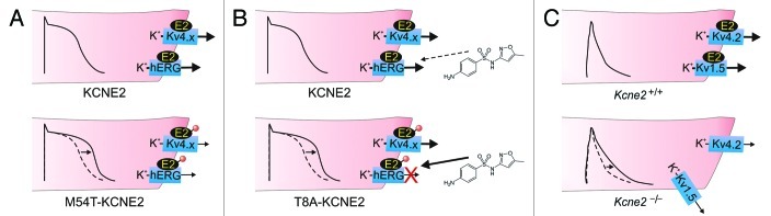
KCNE2, originally designated MinK-related peptide 1 (MiRP1), belongs to a five-strong family of potassium channel ancillary (β) subunits that, despite the diminutive size of the family and its members, has loomed large in the field of ion channel physiology. KCNE2 dictates K (+) channel gating, conductance, α subunit composition, trafficking and pharmacology, and also modifies functional properties of monovalent cation-nonselective HCN channels. The Kcne2 (-/-) mouse exhibits cardiac arrhythmia and hypertrophy, achlorhydria, gastric neoplasia, hypothyroidism, alopecia, stunted growth and choroid plexus epithelial dysfunction, illustrating the breadth and depth of the influence of KCNE2, mutations which are also associated with human cardiac arrhythmias. Here, the modus operandi and physiological roles of this potent regulator of membrane excitability and ion secretion are reviewed with particular emphasis on the ability of KCNE2 to shape the electrophysiological landscape of both excitable and non-excitable cells.

Keywords: KCNQ1; MiRP1; cardiac arrhythmia; choroid plexus; gastric acid; hypothyroidism; thyroid.


Figures

None
Figure 1. KCNE2 topology and stoichiometry. (A) Transmembrane topology (left) and potential stoichiometries (right) of KCNE2 in a Kv channel complex with four Kv α subunits. (B) KCNE2 can control multiple facets of the life of a K+ channel complex. (1) KCNE2 can travel to the plasma membrane in the absence of α subunits and (2) potentially be secreted outside the cell, whereas KCNE1 and KCNE3 appear to require α subunits for anterograde trafficking (3). (4) KCNE2 can prevent N-type α subunits from reaching the plasma membrane, an inhibition released by same-subfamily delayed rectifer α subunits (5). (6) KCNE2 can direct α subunit targeting in polarized cells differentially to, e.g., KCNE3 (7), and KCNE2 can alter multiple functional aspects of surface expressed α-KCNE2 complexes (8). KCNE2 can also mediate α subunit internalization from the plasma membrane (9). References in text.
None
Figure 2. KCNE2 in epithelial cells. (A) KCNE2 in the gastric epithelium. Left, organization of an oxyntic gland in the stomach, with the parietal cell highlighted. Center, the gastric H+/K+-ATPase requires a luminal K+ recycling pathway for gastric acidification, formed by KCNQ1 (Q1) and KCNE2 (E2). Right, in Kcne2−/− mice, KCNQ1 is incorrectly targeted to the parietal cell basolateral membrane by upregulated KCNE3 (E3), and the mice are achlorydric because the gastric H+/K+-ATPase lacks a suitable K+ recycling pathway. (B) KCNE2 in the choroid plexus epithelium. Left, organization of the choroid plexus epithelium (CPe). Center, KCNE2 forms apical K+ channel complexes with KCNQ1 and Kv1.3, which probably contribute to setting membrane potential and regulating anion secretion into the CSF. Right, in Kcne2−/− mice, KCNQ1 and Kv1.3 are incorrectly targeted to the CPe cell basolateral membrane where they pass abnormally large outward K+ currents. Anion secretion appears to be increased, probably because the larger outward K+ current hyperpolarizes the CPe cell Em. (C) KCNE2 in the thyroid epithelium. Left, organization of a thyroid follicle, skirted by thyroid epithelial cells (thryocytes). Center, biosynthesis of thyroid hormones T3 and T4 requires I- to pass from the blood into the central colloid, where it is oxidized and organified by incorporation into thyroglobulin (iodination and conjugation). The product is endocytosed back into the thyrocyte, converted to T3 and T4 by proteolysis, then transported into the blood. KCNQ1-KCNE2 channels form on the basolateral membrane, as does the sodium iodide symporter (NIS). Right, in Kcne2−/− mice (and in Kcnq1−/− mice), thyroid hormone biosynthesis is disrupted by an incompletely understood mechanism characterized by impaired thyroid I- accumulation.
None
Figure 3. KCNE2 in ventricular myocytes. (A) KCNE2 is thought to regulate hERG and Kv4.x channels in the ventricular cardiomyocytes of large mammals. Inherited mutations (red circle) in human KCNE2 that cause loss of function of hERG-KCNE2 channels (and also in some cases Kv4.x-KCNE2 complexes) are associated with delayed ventricular repolarization manifesting clinically as Long QT syndrome. This probably arises from prolonged ventricular action potentials (arrow) arising from reduced K+ flux, especially in phase 3. One example is M54T-KCNE2, a mutation that speeds hERG-KCNE2 channel deactivation. (B) Some human KCNE2 polymorphisms, including T8A, have no appreciable effect on channel function at baseline, but increase the susceptibility of hERG-KCNE2 channels to inhibition by therapeutic agents, in this case sulfamethoxazole, a component of the antibiotic Bactrim. This inhibition can lengthen the QT interval and predispose to drug-induced torsades de pointes, a dangerous ventricular arrhythmia. (C) In mice, KCNE2 regulates Kv4.2 and Kv1.5 in ventricular myocytes. Kcne2 deletion reduces Kv4.2 and Kv1.5 currents and increases mouse ventricular myocyte action potential duration (arrow). Kv1.5 targeting to the intercalated discs is also impaired in Kcne2−/− mouse ventricles.

References

    1. Catterall WA. Structure and function of voltage-gated ion channels. Annu Rev Biochem. 1995;64:493–531. doi: 10.1146/annurev.bi.64.070195.002425. - DOI - PubMed
    1. Goldstein SA, Bockenhauer D, O'Kelly I, Zilberberg N. Potassium leak channels and the KCNK family of two-P-domain subunits. Nat Rev Neurosci. 2001;2:175–84. doi: 10.1038/35058574. - DOI - PubMed
    1. Barry DM, Nerbonne JM. Myocardial potassium channels: electrophysiological and molecular diversity. Annu Rev Physiol. 1996;58:363–94. doi: 10.1146/annurev.ph.58.030196.002051. - DOI - PubMed
    1. Dedek K, Waldegger S. Colocalization of KCNQ1/KCNE channel subunits in the mouse gastrointestinal tract. Pflugers Archiv. 2001;442:896–902. doi: 10.1007/s004240100609. - DOI - PubMed
    1. Song P, Groos S, Riederer B, Feng Z, Krabbenhoft A, Smolka A, et al. KCNQ1 is the luminal K+ recycling channel during stimulation of gastric acid secretion. J Physiol. 2009;587:3955–65. doi: 10.1113/jphysiol.2009.173302. - DOI - PMC - PubMed

Publication types

MeSH terms