. 2012 Apr 18;32(16):5620-30.
doi: 10.1523/JNEUROSCI.0030-12.2012.

Regulation of neuronal mRNA translation by CaM-kinase I phosphorylation of eIF4GII

Affiliations

Regulation of neuronal mRNA translation by CaM-kinase I phosphorylation of eIF4GII

Taasin Srivastava et al. J Neurosci. .

Abstract

Ca²⁺/calmodulin-dependent kinases (CaMKs) are essential for neuronal development and plasticity, processes requiring de novo protein synthesis. Roles for CaMKs in modulating gene transcription are well established, but their involvement in mRNA translation is evolving. Here we report that activity-dependent translational initiation in cultured rat hippocampal neurons is enhanced by CaMKI-mediated phosphorylation of Ser1156 in eukaryotic initiation factor eIF4GII (4GII). Treatment with bicuculline or gabazine to enhance neuronal activity promotes recruitment of wild-type 4GII, but not the 4GII S1156A mutant or 4GI, to the heterotrimeric eIF4F (4F) complex that assembles at the 5' cap structure (m⁷GTP) of mRNA to initiate ribosomal scanning. Recruitment of 4GII to 4F is suppressed by pharmacological inhibition (STO-609) of CaM kinase kinase, the upstream activator of CaMKI. Post hoc in vitro CaMKI phosphorylation assays confirm that activity promotes phosphorylation of S1156 in transfected 4GII in neurons. Changes in cap-dependent and cap-independent translation were assessed using a bicistronic luciferase reporter transfected into neurons. Activity upregulates cap-dependent translation, and RNAi knockdown of CaMKIβ and γ isoforms, but not α or δ, led to its attenuation as did blockade of NMDA receptors. Furthermore, RNAi knockdown of 4GII attenuates cap-dependent translation and reduces density of dendritic filopodia and spine formation without effect on dendritic arborization. Together, our results provide a mechanistic link between Ca²⁺ influx due to neuronal activity and regulation of cap-dependent RNA translation via CaMKI activation and selective recruitment of phosphorylated 4GII to the 4F complex, which may function to regulate activity-dependent changes in spine density.


Figures
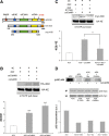
Figure 1.
Figure 1.
CaMKI-mediated phosphorylation of S1156 in eIF4GII (4GII) promotes its recruitment to the eIF4F (4F) initiation complex. A, Schematic showing full-length 4GII (1–1585 aa) with the different protein interaction motifs and the CaMKI phosphorylation site S1156 highlighted. Also shown are the two epitope-tagged partial 4GII constructs used in this study, HA-4GII (140–1585 aa) and Myc-4GII (590–1450 aa)—importantly, both contain the main 4F interaction motifs. B, HEK293 cells were cotransfected with HA-4GII and HA-eIF4E (HA-4E) and, as indicated, with either constitutive active (ca) CaMKK or CaMKI plasmids. After 24 h of expression, lysates were prepared and subjected to m7GTP Sepharose pull-down to isolate the eIF4F (4F) complex (see Materials and Methods). Representative blots probed with anti-HA antibody showing 4GII and 4E associated with m7GTP beads from each experimental condition (top) and quantification of results (bottom, n = 4 independent experiments). Note the increase in 4GII levels, normalized to 4E levels, in the 4F complex from the samples expressing caCaMKK or caCaMKI. C, To test the functional significance of the predicted CaMKI phosphorylation site (S1156), HEK293 cells were transfected as indicated with caCaMKI in combination either with myc-tagged wild-type 4GII (4GII) or S1156A 4GII and subject to m7GTP Sepharose pull-down as in B. Top, Representative blots probed with anti-myc or anti-HA antibodies, respectively, showing the wild-type 4GII and S1156A 4GII associated with the 4F complex. Bottom, Quantification of wild-type and S1156A 4GII in the 4F complex from each condition (n = 4 independent experiments). D, Activity-dependent S1156 phosphorylation in cultured hippocampal neurons. Hippocampal neurons were transfected at DIV7 to express myc-tagged wild-type 4GII or 4GII-S1156A plasmids. At DIV12, neurons were left untreated or stimulated with either 20 μm bicuculline (Bic) or 10 μm gabazine (GABAz) for 45 min. Lysates were prepared, 4GII was immunoprecipitated with anti-Myc antibody and then an in vitro kinase assay was performed in the presence of purified caCaMKI (see Materials and Methods). Top, Representative autoradiographs depicting in vitro CaMKI-mediated back-phosphorylation in the presence of [γ-32P]ATP. Middle, Representative blots probed with anti-myc antibody showing the immunoprecipitated (IP) 4GII and total cell lysate (tcl) 4GII protein in neurons after 5 d of expression. Bottom, Quantification of autoradiographs (arbitrary units, a.u.) for the conditions indicated (n = 2 independent experiments). Error bars indicate SEM; where indicated, ***p < 0.001, *p < 0.05 by Student's t test.
Figure 2.
Figure 2.
Neuronal activity enhances 4GII recruitment to the 4F complex in primary hippocampal neurons. DIV12–14 primary hippocampal neurons were either left untreated (controls) or treated with 16 mm KCl, 20 μm bicuculline (Bic) or 10 μm gabazine (GABAz) for the indicated time periods. Lysates were subject to the m7GTP Sepharose pull down and the corresponding blots were probed with anti-eIF4GII (4GII), anti-eIF4A (4A) or anti-eIF4E (4E) antibodies. A–C, Representative blots show the increase in endogenous 4GII recruitment to the 4F complex following KCl or Bic (A), or gabazine treatments (B). Quantification of 4GII recruited to the 4F complex following induction of neuronal activity (C) (n = 4 independent experiments). D, Representative blot (left panel) and quantification (right panel, n = 4) showing no change in total protein levels for 4GII following Bic treatment. Total ERK2 was used as a loading control. Error bars indicate SEM; ***p < 0.001 by Student's t test compared with untreated controls.
Figure 3.
Figure 3.
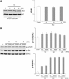
Effects of neuronal activity on 4GI recruitment to the 4F complex and activation of mTOR and ribosomal S6 kinase. A, DIV12–14 hippocampal neurons were left untreated (control) or treated with 20 μm bicuculline (Bic) for the indicated times before isolation of the 4F complex by m7GTP Sepharose pull down. Representative blot (left) and quantification (right, n = 4) show recruitment of 4GI, in contrast to 4GII, to the 4F complex is not affected by neuronal activity. B, Effects of neuronal activity on mTOR activation and ribosomal S6 phosphorylation. DIV12–14 hippocampal neurons were either left untreated (control) or treated with 20 μm Bic or 10 μm gabazine (GABAz) for the indicated time points. Left, representative blots showing activation of mTOR kinase (phospho-Ser2448) and rS6 protein (phospho-Ser235/236) following activity. Note, the modest (p > 0.05) activation of mTOR kinase and hyper-activation and phosphorylation of S6 protein. Total ERK2 was used as loading control. Right, quantification of mTOR kinase activity (top) and ribosomal S6 protein (bottom) phosphorylation normalized to unstimulated controls (n = 3 independent experiments). Blots were probed with primary phospho-specific anti-mTOR (p-mTOR) and anti-S6 (p-S6) as well as anti-ERK2 antibodies where indicated. Error bars indicate SEM.
Figure 4.
Figure 4.
4GII recruitment to the 4F complex is dependent on calcium and CaMKK signaling in hippocampal neurons. A, DIV12–14 hippocampal neurons were left untreated (control) or treated with or without 20 μm BAPTA-AM for 30 min to chelate calcium followed by 20 μm bicuculline (Bic) treatment for 45 min. The left panel illustrates representative blots whereas the right panel shows quantification (n = 4 independent experiments). B, 4GII recruitment to the 4F complex is dependent on CaMKK and neuronal activity. DIV12–14 neurons were either (1) untreated (control) or treated with 20 μm muscimol or 20 μm bicuculline (Bic), alone for 45 min, or (2) treated with 1 μm TTX for 1 h, or (3) pretreated with 50 μm STO-609 (CaMKK inhibitor) for 4 h followed by Bic treatment for 45 min (n = 4 independent experiments). C, Bicuculline activation of CaMKI (pCaMKI) is blocked by the CaMKK inhibitor STO-609. Experimental conditions are as in B (n = 4). Representative blots for pCaMKI (left) and quantification (n = 4 independent experiments, right) are shown. Total ERK2 was used as a loading control. Error bars indicate SEM; where indicated, ***p < 0.001 by Student's t test.
Figure 5.
Figure 5.
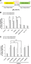
Activity-mediated translation in hippocampal neurons is dependent on NMDAR and CaMKI signaling. A, Schematic of the pRL-HCV-FL bicistronic luciferase reporter. The Renilla Luciferase (RLuc) gene is under the control of the m7GTP cap, hence it is cap-dependent. The Firefly Luciferase (FLuc) gene is under the control of IRES element derived from the 5′UTR of HCV to make it cap-independent. B, DIV10–11 hippocampal neurons were transfected with pRL-HCV-FL and after 24 h of expression were either left untreated (controls) or treated for 24 h with 16 mm KCl, 20 μm bicuculline (Bic, alone or in combination with 1 μm TTX), 10 μm gabazine (GABAz, alone or in combination with 50 μm AP-5 or 3 μm Ro25). The activity from the reporter genes (RLuc and FLuc) was then quantified by dual luciferase assays (see Materials and Methods). Neuronal activity preferentially enhances cap-dependent translation of RLuc (cap-dependent) over the FLuc levels (cap-independent) (n = 4 independent experiments). C, DIV10–11 neurons were cotransfected with pRL-HCV-FL either with vector or with various shRNA constructs to knockdown the different CaMKI isoforms (α, β, γ, or δ; a, b, g, d, respectively) or transfected with CaMKIIN to inhibit CaMKII. After 24 h of transfection, the neurons were either left untreated (control) or treated with 10 μm gabazine for an additional 24 h. Activity-dependent cap-dependent translation, but not cap-independent translation, was sensitive to knockdown of CaMKIγ and CaMKIβ. In contrast, knockdown of CaMKIα, CaMKIδ, or CaMKII had no significant effect on reporter gene expression (n = 4 independent experiments). Error bars indicate SEM; where indicated, **p < 0.01, ***p < 0.001 by Student's t test.
Figure 6.
Figure 6.
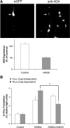
4GII plays a role in initiation of cap-dependent translation in hippocampal neurons in an activity-dependent manner. A, DIV10–11 neurons cotransfected for 3 d with shRNA eIF4GII plasmid coexpressing eGFP (sh4GII) (to identify transfected neurons) were fixed and analyzed for effective knockdown by immunofluorescence using primary anti-eIF4GII antibody (see Materials and Methods). Top, Representative immunofluorescence images showing knockdown of 4GII expression in a shRNA transfected neuron (arrow) whereas expression in the nontransfected neurons is unaffected. Bottom, Quantification and comparison of 4GII expression in neurons with and without shRNA knockdown (n = 4 coverslips per condition; n = 2 independent experiments). B, DIV10–11 neurons were cotransfected with pRL-HCV-FL with or without sh4GII for 3 d and then treated with 10 μm gabazine (GABAz) as described in panel 5B. Knockdown of 4GII specifically attenuated RLuc (cap-dependent) expression (n = 4 independent experiments). Error bars indicate SEM; where indicated, *p < 0.05 by Student's t test.
Figure 7.
Figure 7.
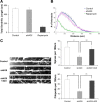
Knockdown of 4GII leads to a decrease in density of dendritic filopodia and spines. DIV5 neurons were transfected with mRFP alone and left (1) untreated (controls) or treated with 1 μm rapamycin where indicated or (2) cotransfected in combination with shRNA eIF4GII (sh4GII) and empty vector or Myc-4GII (590–1450). A, B, Neurons were fixed at DIV12 and their dendrites were imaged and analyzed (see Materials and Methods). A, Bar graphs illustrating no significant change in total dendritic lengths compared with controls following 4GII knockdown but strong suppression by rapamycin. B, Sholl analysis, illustrating minimal effects on overall dendritic arborization following knockdown of 4GII. Rapamycin treatment, in contrast to 4GII suppression, has dramatic effects on both total dendritic lengths and dendritic arborization (A, B). C, Knockdown of 4GII strongly suppresses spine and filopodia density. Left, High-resolution images of the dendrites from DIV12 neurons depicting protrusions from a representative neuron from each indicated experimental condition. One proximal dendrite at least 100 μm in length per neuron from each experimental condition was analyzed for both spine and filopodia protrusions (n = 2–3 neurons per coverslip; 4 coverslips per condition from 3 independent experiments). Right, Knockdown of 4GII has an effect on both spine and filopodia density, and these effects are rescued by coexpression of Myc-4GII, which is insensitive to the sh4GII. Error bars indicate SEM; where indicated, **p < 0.01, *p < 0.05 by Student's t test.

References

    1. Atkins CM, Nozaki N, Shigeri Y, Soderling TR. Cytoplasmic polyadenylation element binding protein-dependent protein synthesis is regulated by calcium/calmodulin-dependent protein kinase II. J Neurosci. 2004;24:5193–5201. - PMC - PubMed
    1. Beretta L, Gingras AC, Svitkin YV, Hall MN, Sonenberg N. Rapamycin blocks the phosphorylation of 4E-BP1 and inhibits cap-dependent initiation of translation. EMBO J. 1996;15:658–664. - PMC - PubMed
    1. Bramham CR, Wells DG. Dendritic mRNA: transport, translation and function. Nat Rev Neurosci. 2007;8:776–789. - PubMed
    1. Caron S, Charon M, Cramer E, Sonenberg N, Dusanter-Fourt I. Selective modification of eukaryotic initiation factor 4F (eIF4F) at the onset of cell differentiation: recruitment of eIF4GII and long-lasting phosphorylation of eIF4E. Mol Cell Biol. 2004;24:4920–4928. - PMC - PubMed
    1. Cavazzini M, Bliss T, Emptage N. Ca2+ and synaptic plasticity. Cell Calcium. 2005;38:355–367. - PubMed

Publication types

MeSH terms

Substances