. 2012 Apr 18;32(16):5654-66.
doi: 10.1523/JNEUROSCI.0455-12.2012.

Neurogenic subventricular zone stem/progenitor cells are Notch1-dependent in their active but not quiescent state

Affiliations

Neurogenic subventricular zone stem/progenitor cells are Notch1-dependent in their active but not quiescent state

Onur Basak et al. J Neurosci. .

Abstract

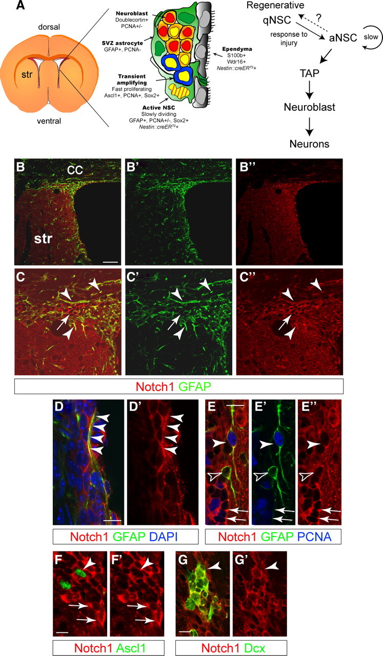
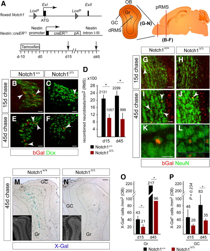
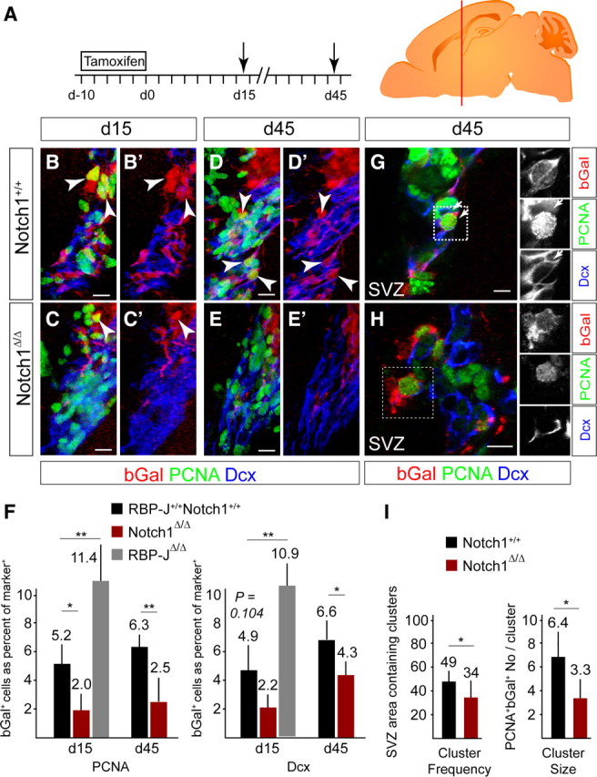
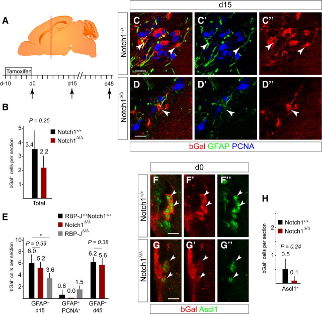
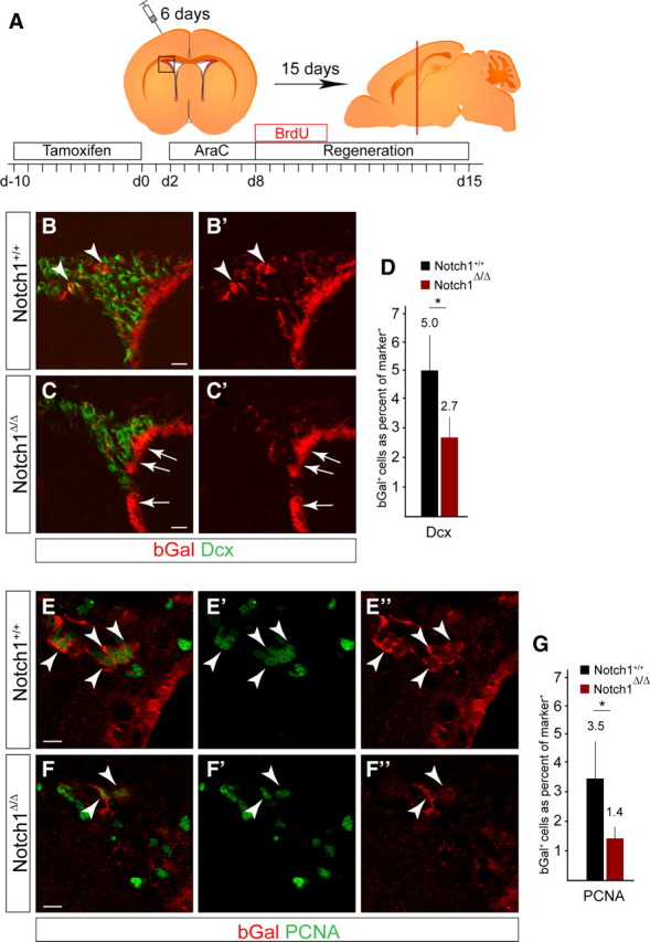
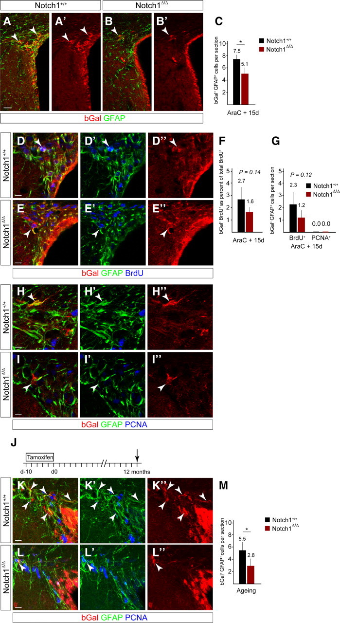
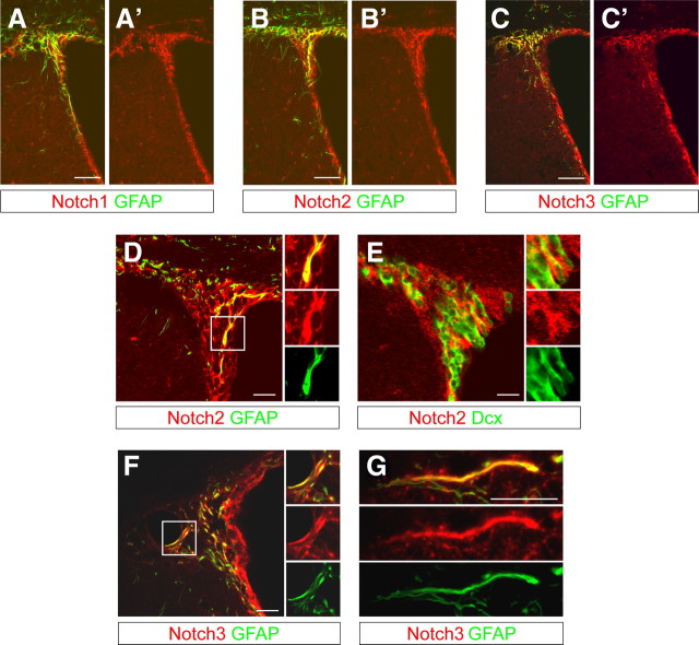
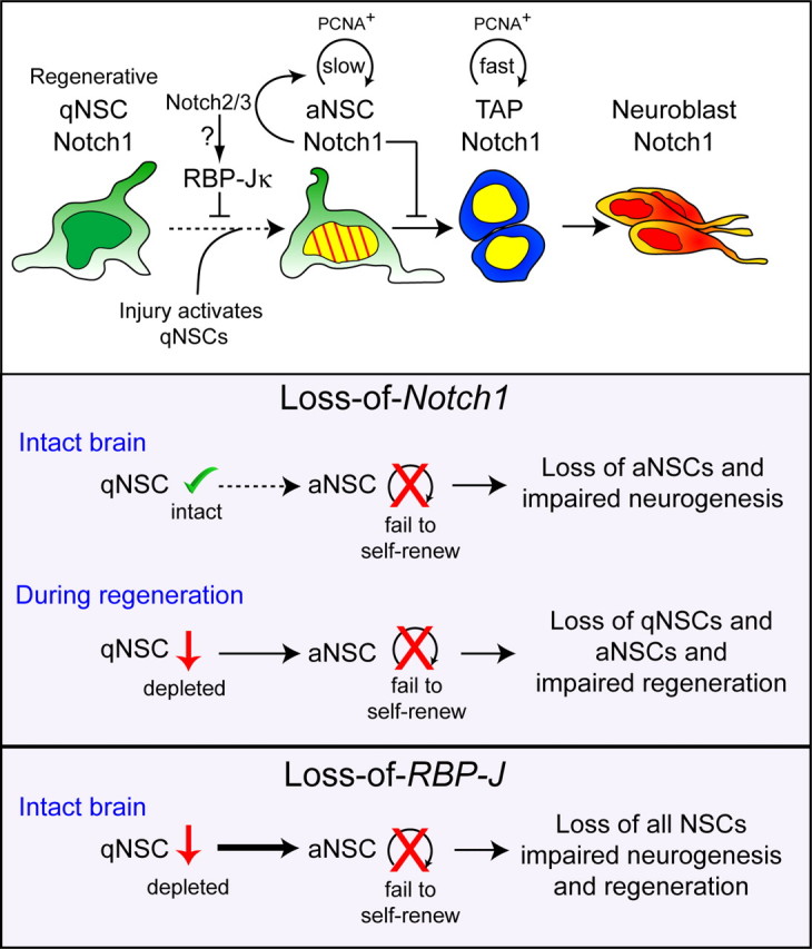
The adult mammalian forebrain contains neural stem/progenitor cells (NSCs) that generate neurons throughout life. As in other somatic stem cell systems, NSCs are proposed to be predominantly quiescent and proliferate only sporadically to produce more committed progeny. However, quiescence has recently been shown not to be an essential criterion for stem cells. It is not known whether NSCs show differences in molecular dependence based on their proliferation state. The subventricular zone (SVZ) of the adult mouse brain has a remarkable capacity for repair by activation of NSCs. The molecular interplay controlling adult NSCs during neurogenesis or regeneration is not clear but resolving these interactions is critical in order to understand brain homeostasis and repair. Using conditional genetics and fate mapping, we show that Notch signaling is essential for neurogenesis in the SVZ. By mosaic analysis, we uncovered a surprising difference in Notch dependence between active neurogenic and regenerative NSCs. While both active and regenerative NSCs depend upon canonical Notch signaling, Notch1-deletion results in a selective loss of active NSCs (aNSCs). In sharp contrast, quiescent NSCs (qNSCs) remain after Notch1 ablation until induced during regeneration or aging, whereupon they become Notch1-dependent and fail to fully reinstate neurogenesis. Our results suggest that Notch1 is a key component of the adult SVZ niche, promoting maintenance of aNSCs, and that this function is compensated in qNSCs. Therefore, we confirm the importance of Notch signaling for maintaining NSCs and neurogenesis in the adult SVZ and reveal that NSCs display a selective reliance on Notch1 that may be dictated by mitotic state.


Figures

Figure 1.
Figure 1.
Notch1 is expressed in the adult subventricular zone niche. A, Schematic view of the SVZ and cell-type specific marker expression (adapted from Doetsch et al., 1999b). Hierarchical organization of the cells involved in adult neurogenesis in the SVZ. aNSC divide infrequently and generate TAPs, which in turn generate neuroblasts. Quiescent regenerative NSCs respond to signals (including injury), enter the cell cycle, and generate neurogenic aNSCs. It is unclear whether aNSC and regenerative qNSCs are independent states of the same cell and whether NSCs can shuttle between the two. Striped yellow nucleus is slow dividing, BrdU-retaining NSC; yellow nucleus is mitotically active cell. B, Low-magnification, confocal optical sections showing overview images of the lateral wall of the forebrain ventricle, including the SVZ. C, Notch1 protein codistributes with GFAP (arrowheads) but is also expressed by GFAP cells (arrow). Higher-magnification images of sections in B. D, GFAP+ SVZ astrocytes express Notch1, including putative NSCs, with processes contacting the lateral ventricle (arrowheads) and interdigitated into the ependymal lining. E, Some astrocytes (GFAP+) are in the cell cycle (PCNA+, arrowheads) and some are not (GFAP+PCNA, open arrowheads). SVZ astrocytes have lower levels of Notch1 compared with clustered putative neuroblasts (arrows). F, Neurogenic TAPs (Ascl1+) express Notch1 (arrowhead) comparable to other SVZ cells (arrows). G, Dcx+ neuroblasts (arrowhead) also express Notch1 (Nyfeler et al., 2005). Str, striatum; CC, corpus callosum. Scale bars: B, 100 μm; D–G, 20 μm.
Figure 2.
Figure 2.
Notch1 ablation reduces the number of neuroblasts in the RMS and the number of newborn neurons in all OB layers. A, Scheme of the floxed Notch1 locus (Exon I flanked with LoxP sites) and Nestin::creERT2 alleles used in the analysis (Giachino and Taylor, 2009). Adult mice were induced with TAM for 10 d, followed by a 15 or 45 d chase (death, arrow). Brains were analyzed on coronal sections at the level of the red bar (OB and proximal RMS). Schematic view of the OB showing the distal RMS (dRMS), granule cell (Gr) and glomerular cell layers (GC). B, C, bGal+Dcx+ neuroblasts (arrowheads) in the proximal RMS (pRMS) are reduced in Notch1 cKO (Notch1Δ/Δ) compared with control (Notch1+/+) animals 15 d after TAM treatment. D, Quantification of bGal+ neuroblasts in the RMS of Notch1 cKO (Notch1Δ/Δ; n = 3) and control (Notch1+/+; n = 4) animals 15 and 45 d after TAM treatment. The production of bGal+ neuroblasts is reduced by day 15 and does not recover in Notch1 cKO animals. E, F, bGal+Dcx+ neuroblasts (arrowheads) in the posterior RMS are reduced in Notch1 cKO (Notch1Δ/Δ) compared with control (Notch1+/+) animals 45 d after TAM treatment. G–L, bGal+ cells in the OB of both control (Notch1+/+) and Notch1 cKO (Notch1Δ/Δ) mice are NeuN+ neurons (arrowheads). M, N, Ablation of Notch1 dramatically reduces the number of newly generated neurons (X-Gal stained blue) in the OB 45 d after TAM treatment. Inserts shows DAPI staining of the corresponding sections. O, P, Quantification of recombined cells in the granule cell (O) and glomerular cell layers (P) of the OB of control (Notch1+/+; n = 3 and 8, respectively) and Notch1 cKO (Notch1Δ/Δ; n = 3 and 6, respectively) mice. Error bars are SD. Student's t test, *p < 0.05. Scale bars: B, C, E–G, I, K, 20 μm; M, N, 100 μm.
Figure 3.
Figure 3.
Notch1 ablation results in a persistent loss of progenitors in the SVZ. A, Scheme of the TAM induction protocol in adult mice followed by a 15 or 45 d chase (death, arrow). Brains were analyzed on coronal sections of the SVZ at the level of the red bar. B, C, bGal+PCNA+ proliferating cells (arrowheads) and neuroblasts (bGal+Dcx+) are lost in the SVZ of Notch1 cKO mice (Notch1Δ/Δ; C) compared with controls (Notch1+/+; B) 15 d after TAM treatment. D, E, Loss of proliferating cells (bGal+PCNA+; arrowheads) and neuroblasts (bGal+Dcx+) in the SVZ of Notch1 cKO mice (Notch1Δ/Δ; E) compared with controls (Notch1+/+; D) even 45 d after the recombination. F, Quantification of bGal+PCNA+ proliferating cells and neuroblasts in the SVZ of Notch1 cKO (Notch1Δ/Δ; n = 3–4), RBP-J cKO (RBP-JΔ/Δ; n = 3), and control (RBP-J+/+Notch1+/+; n = 3–4) animals. G, Sparse recombination enables cell-autonomous cluster analysis of the progeny of Nestin::creERT2-expressing NSCs (bGal+) including proliferating cells (bGal, PCNA+; arrows) in the SVZ. H, bGal+ neuroblasts [Doublecortin (Dcx+)] in the RMS are clustered, consistent with having a common origin. I, Quantification of clusters of recombined proliferating cells and the number of bGal+PCNA+ cells per cluster in Notch1 cKO (Notch1Δ/Δ; n = 6) and control (Notch1+/+; n = 7) animals. Probability of recombined clusters per 6.25 × 105 μm3 of the SVZ. Error bars are SD. Student's t test, *p < 0.05, **p < 0.001. Scale bars, 20 μm.
Figure 4.
Figure 4.
Notch1 ablation does not result in a loss of GFAP+ NSC. A, Ten day TAM induction protocol in adult mice followed by 0, 15, or 45 d chase (death, arrow). Brains were analyzed on coronal sections at the level of the red bar. B, Ablation of Notch1 did not significantly affect the total number of bGal+ cells at day 0. Notch1 cKO (Notch1Δ/Δ; n = 3) and control (Notch1+/+; n = 3) animals. C, Consistent with targeting early progenitors, a minor cell population are bGal+ in the subependymal layer of the SVZ in control animals (Notch1+/+). Most of the targeted cells (bGal+) that express the stem cell marker GFAP are quiescent (PCNA, arrowheads). D, bGal+ cells in the subependymal layer of the SVZ in Notch1 cKO animals (Notch1Δ/Δ) still express GFAP (arrowhead). E, Notch1 ablation does not alter the number of bGal+ cells that expressed GFAP+ in the subependymal layer of the SVZ and proliferation (PCNA) of bGal+GFAP+ cells was also not increased. In contrast, RBP-J ablation (RBP-JΔ/Δ) results in reduced bGal+GFAP+ cells in the subependymal layer. Notch1 cKO (Notch1Δ/Δ; n = 3 at day 15 and 4 at day 45), RBP-J cKO (RBP-JΔ/Δ; n = 3), and control (RBP-J+/+Notch1+/+; n = 7 at day 15 and 4 at day 45) animals. F, bGal+ cells rarely express the TAP marker Ascl1 in control mice immediately after TAM treatment (day 0: Notch1+/+; arrowheads). G, Ascl1+ recombined cells are detectable in Notch1 cKO mice and are not increased (Notch1Δ/Δ, arrowheads). H, Ablation of Notch1 did not significantly affect the number of bGal+Ascl1+ TAPs at d0. Notch1 cKO (Notch1Δ/Δ, n = 3) and control (Notch1+/+, n = 3) animals. Error bars are SD. Student's t test, *p < 0.05. Scale bars, 10 μm.
Figure 5.
Figure 5.
Notch1 is required for regeneration of neurogenic progenitors in the SVZ. A, Schematic overview of the experimental paradigm and regenerative process. Notch1lox/lox and control littermates were induced with TAM for 10 d before intracranial infusion of AraC. Mice were treated with BrdU during the first 5 d of regeneration. Brains were analyzed on coronal sections at the level shown by the red bar. B, bGal+ dormant NSCs activate following AraC-induced degeneration and contribute to regeneration in the SVZ by generating newborn bGal+Dcx+ neuroblasts (arrowheads). C, bGal+ cells are reduced in the regenerated SVZ of Notch1 cKO (Notch1Δ/Δ) animals. The ependymal layer is not affected in the Notch1 cKO even after AraC treatment (arrows). D, Quantification of bGal+ neuroblasts (Dcx+) in the regenerated SVZ of Notch1 cKO (Notch1Δ/Δ; n = 4) and control (Notch1+/+; n = 4) animals. E, bGal+ NSCs generate clusters of proliferating progeny (bGal+PCNA+, arrowheads) in the regenerated SVZ of controls (Notch1+/+). F, bGal+PCNA+ cells are reduced in the regenerated SVZ of Notch1 cKO (Notch1Δ/Δ) animals (arrowheads). G, Quantification of bGal+ proliferating cells (PCNA+) in the regenerated SVZ of Notch1 cKO (Notch1Δ/Δ; n = 4) and control (Notch1+/+; n = 4) animals. Error bars are SD. Student's t test, *p < 0.05. Scale bars: B, C, 20 μm; E, F, 10 μm.
Figure 6.
Figure 6.
Notch1 ablation results in a reduction in GFAP+ dormant NSCs in the regenerated SVZ and during aging. A, B, bGal+ cells (arrowheads) in the regenerated SVZ 15 d after stopping AraC infusion are reduced in the Notch1-ablated (Notch1Δ/Δ) compared with control (Notch1+/+) animals. C, Ablation of Notch1 (Notch1Δ/Δ; n = 5) before regeneration results in a reduction in bGal+GFAP+ cells compared with control (Notch1+/+; n = 5) animals. D, E, BrdU label-retaining cells are present in the regenerated SVZ of control (Notch1+/+) and Notch1 cKO (Notch1Δ/Δ) animals and some express GFAP (arrowheads). F, bGal+ BrdU-retaining cells are slightly reduced in the regenerated SVZ following deletion of Notch1 (Notch1Δ/Δ; n = 5) compared with control (Notch1+/+; n = 5) animals. G, The number of bGal+GFAP+ BrdU-retaining cells is slightly reduced in the regenerated SVZ of Notch1 cKO (Notch1Δ/Δ; n = 5) compared with control (Notch1+/+; n = 5) animals. bGal+GFAP+ cells in the Notch1 cKO SVZ do not remain mitotically active (PCNA+) after regeneration. H, I, In both control and Notch1 cKO animals, GFAP+ cells return to a quiescent state (PCNA) in the regenerated SVZ (arrowheads). J, 10 d TAM induction protocol in adult mice followed by 12-month chase (death, arrow). K–M, Ablation of Notch1 in young adult mice (Notch1Δ/Δ; n = 4) results in a reduction in bGal+GFAP+ cells when analyzed 1 year later compared with control mice (Notch1+/+; n = 4). Error bars are SD. Student's t test, *p < 0.05. Scale bars: A, 25 μm; D, E, H, I, K, L, 10 μm.
Figure 7.
Figure 7.
Notch2 and Notch3 are expressed in the SVZ with a similar pattern to Notch1. A, Notch1 is expressed in the SVZ of adult mice by GFAP+ and GFAP cells. B, C, Consecutive sections stained with anti-Notch2 (B) or anti-Notch3 (C) antibodies showing a comparable expression pattern in the SVZ to that of Notch1. D, Notch2 is expressed by GFAP cells in the subependymal region of the SVZ but also by GFAP+ SVZ astrocytes and putative NSCs in close proximity to or making contact with the lateral ventricle. E, Some Notch2+GFAP cells are Doublecortin+ (Dcx+) neuroblasts. F, G, Notch3 is prominently expressed by GFAP+ SVZ astrocytes. Scale bars: A–C, 100 μm; D–G, 20 μm.
Figure 8.
Figure 8.
Model of Notch1 regulation of SVZ homeostasis and regeneration. Role of Notch1 in adult NSC populations. Continuous neurogenesis in the adult SVZ is maintained by aNSCs, which divide slowly (striped yellow nucleus). aNSCs depend on Notch1 for maintenance, self-renewal, and neurogenesis; in the absence of Notch1, these aNSCs are compromised. Fast-dividing TAPs (yellow nucleus) express Notch1 and give rise to neuroblasts that migrate to the OB and generate neurons. qNSCs are dependent on RBP-J but not Notch1 for their maintenance, and enter the cell cycle in response to injury to become the driving force for regeneration. The selective loss of aNSCs following Notch1 deletion suggests an uncompensated role for Notch1 in homeostatic neurogenesis. In the absence of Notch1, activated NSCs fail to self-renew and effectively reinstate adult neurogenesis, resulting in a reduction of both qNSCs and a loss of aNSCs. qNSCs do not readily transit between states in the intact brain but during regeneration, qNSCs enter the cell cycle and assume an aNSC Notch1-dependent state. RBP-J blocks cell cycle entry of qNSCs and promotes aNSC maintenance. Slow/fast: Rate of cell division.

References

    1. Ables JL, Decarolis NA, Johnson MA, Rivera PD, Gao Z, Cooper DC, Radtke F, Hsieh J, Eisch AJ. Notch1 is required for maintenance of the reservoir of adult hippocampal stem cells. J Neurosci. 2010;30:10484–10492. - PMC - PubMed
    1. Aguirre A, Rubio ME, Gallo V. Notch and EGFR pathway interaction regulates neural stem cell number and self-renewal. Nature. 2010;467:323–327. - PMC - PubMed
    1. Balordi F, Fishell G. Mosaic removal of hedgehog signaling in the adult SVZ reveals that the residual wild-type stem cells have a limited capacity for self-renewal. J Neurosci. 2007;27:14248–14259. - PMC - PubMed
    1. Basak O, Taylor V. Identification of self-replicating multipotent progenitors in the embryonic nervous system by high Notch activity and Hes5 expression. Eur J Neurosci. 2007;25:1006–1022. - PubMed
    1. Basak O, Taylor V. Stem cells of the adult mammalian brain and their niche. Cell Mol Life Sci. 2009;66:1057–1072. - PMC - PubMed

Publication types

MeSH terms

LinkOut - more resources