. 2012 Jun;86(12):6835-46.
doi: 10.1128/JVI.00437-12. Epub 2012 Apr 18.

Initial HIV-1 antigen-specific CD8+ T cells in acute HIV-1 infection inhibit transmitted/founder virus replication

Affiliations

Initial HIV-1 antigen-specific CD8+ T cells in acute HIV-1 infection inhibit transmitted/founder virus replication

Stephanie A Freel et al. J Virol. 2012 Jun.

Abstract

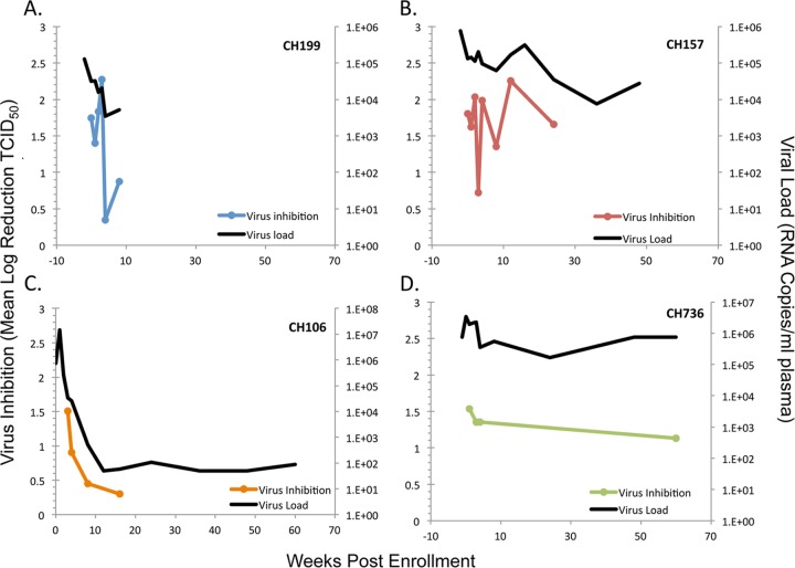
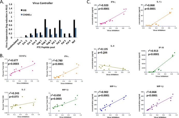
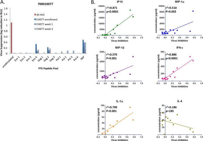
CD8-mediated virus inhibition can be detected in HIV-1-positive subjects who naturally control virus replication. Characterizing the inhibitory function of CD8(+) T cells during acute HIV-1 infection (AHI) can elucidate the nature of the CD8(+) responses that can be rapidly elicited and that contribute to virus control. We examined the timing and HIV-1 antigen specificity of antiviral CD8(+) T cells during AHI. Autologous and heterologous CD8(+) T cell antiviral functions were assessed longitudinally during AHI in five donors from the CHAVI 001 cohort using a CD8(+) T cell-mediated virus inhibition assay (CD8 VIA) and transmitted/founder (T/F) viruses. Potent CD8(+) antiviral responses against heterologous T/F viruses appeared during AHI at the first time point sampled in each of the 5 donors (Fiebig stages 1/2 to 5). Inhibition of an autologous T/F virus was durable to 48 weeks; however, inhibition of heterologous responses declined concurrent with the resolution of viremia. HIV-1 viruses from 6 months postinfection were more resistant to CD8(+)-mediated virus inhibition than cognate T/F viruses, demonstrating that the virus escapes early from CD8(+) T cell-mediated inhibition of virus replication. CD8(+) T cell antigen-specific subsets mediated inhibition of T/F virus replication via soluble components, and these soluble responses were stimulated by peptide pools that include epitopes that were shown to drive HIV-1 escape during AHI. These data provide insights into the mechanisms of CD8-mediated virus inhibition and suggest that functional analyses will be important for determining whether similar antigen-specific virus inhibition can be induced by T cell-directed vaccine strategies.


Figures

Fig 1
Fig 1
CD8+ T cells from HIV-1 donors before or during the time of seroconversion suppress T/F viruses at levels similar to suppression in elite controllers. CD8+ T cells isolated from 3 seronegative donors, 5 HIV-1-positive donors (Fiebig score of ≤5), and 5 HIV-1-positive elite controllers were tested for antiviral activity against a panel of 7 T/F IMC in the CD8 VIA. Dots represent the log reduction in virus replication (RLU) for each CD8 donor against a single virus at an E/T ratio of 2:1 compared to the results from a CD8-depleted infection control. Statistical significance was determined by using the mean virus inhibition across T/F viruses for each donor (Student's t test). The dotted line indicates a log inhibition cutoff for positivity of 0.96 (3 standard deviations above the mean seronegative donor T/F virus inhibition).
Fig 2
Fig 2
CD8+ T cell-mediated virus inhibition of heterologous T/F viruses is dynamic during the acute phase of infection. Antiviral activity was measured from CD8+ T cells isolated longitudinally from 4 HIV-1-positive patients (3 ART-naïve and CH106 who began ART at study week 1) during acute infection (shown as log reduction in virus titer [TCID50] from the results for a CD8-depleted control). Mean inhibition across T/F viruses is shown for 4 patients: (A) 700010199; (B) 702010157; (C) 702010736; (D) 700010106.
Fig 3
Fig 3
CD8+ T cell-mediated inhibition of autologous virus is maintained even in the absence of high viral loads. Antiviral activities exerted by CD8+ T cells that were isolated longitudinally from a clade B-infected HIV-1-positive patient during acute infection are shown as log reductions in titer (TCID50) compared to the results for the CD8 T cell-depleted control sample. CD8+ T cell-mediated inhibition of 3 heterologous T/F viruses (CH040.c, CH058.c, and CH077.t), one autologous T/F virus (CH106.c), and NL4-3 is shown over 48 weeks for patient 7000010106.
Fig 4
Fig 4
Virus rapidly escapes from CD8-driven pressure exerted early after infection. (A) CD8+ T cells isolated from 3 early time points (enrollment to week 3) were tested in the CD8 VIA for inhibitory capacity against autologous (CH077.t) and heterologous (CH058.c) T/F and 6-month viruses. Inhibition of virus replication (TCID50) relative to the results for a CD8-depleted control is shown. (B) Inhibition of T/F and paired 6-month virus by CD8+ T cells from heterologous acute patients is shown as log reduction in virus titer (TCID50) relative to the results for a CD8-depleted control. The range of inhibition for heterologous donors is shown as log reduction in virus titer at 2:1 E/T.
Fig 5
Fig 5
Soluble inhibition of HIV-1 correlates with cytokine expression in CD8+ T cells from a virus controller. Transwell CD8 VIA was used to assess the soluble-factor inhibitory capacity of PTE peptide-stimulated CD8+ T cells from a virus controller. Suppression of virus replication is shown as log reduction in RLU from the results for an infected TZM-bl control. Background is determined using peptide pool-stimulated CD8+ T cells from a seronegative donor and has been subtracted. (A) Suppression of CH040.c T/F and IIIB viruses by peptide pool-stimulated VC9 CD8+ T cells is shown. (B) Each panel shows the percentage of total CD8+ T cells expressing the indicated cytokine of CD107a as determined by flow cytometric analysis. (C) Each panel shows supernatant cytokine levels as measured by Luminex following peptide stimulation of CD8+ T cells and coculture with infected CD4+ T cell targets. Lines indicate best fit, by least squares method. Pearson's correlations and P values are shown (Prism software).
Fig 6
Fig 6
CD8+ T cells inhibit replication of T/F viruses through antigen-specific release of soluble inhibitors. Transwell CD8 VIA assay was used for longitudinal assessment of the soluble-factor inhibitory capacity of HIV-1 peptide-stimulated CD8+ T cells from 4 acute donors. Suppression of virus replication is shown as log reduction in RLU from the results for an infected TZM-bl control. Results for the JR-HVS CD8+ T cell line positive control are shown in red. Background is determined using peptide pool-stimulated CD8+ T cells from a seronegative donor and has been subtracted. (A) Soluble-factor suppression of CH040.c virus by 700010106 CD8+ T cells. (B) Soluble-factor suppression of CH040.c virus by 701010199 CD8+ T cells. (C) Soluble-factor suppression of CH042.c virus by 702010157 CD8+ T cells. (D) Soluble-factor suppression of CH042.c virus by 702010736 CD8+ T cells. (E) A summary of positive responses seen at enrollment or week 1 (cutoff of 2.5 standard deviations above the seronegative mean after background subtraction).
Fig 7
Fig 7
Soluble-factor inhibition of autologous T/F virus corresponds with rapid virus escape and correlates with cytokine release. Transwell CD8 VIA was used for longitudinal assessment of the soluble-factor inhibitory capacity of PTE peptide-stimulated CD8+ T cells from an acute patient. Suppression of virus replication is shown as log reduction in RLU from the results for an infected TZM-bl control. Results for the JR-HVS CD8+ T cell line control are shown in red. Background is determined using peptide pool-stimulated CD8+ T cells from a seronegative donor and was subtracted. (A) Suppression of CH077.t T/F virus by peptide pool-stimulated 700010077 acute CD8+ T cells is shown. (B) Each panel shows supernatant cytokine levels as measured by Luminex following peptide stimulation of CD8+ T cells and coculture with infected CD4+ T cell targets. Pearson's correlations and P values are shown (Prism software).
Fig 8
Fig 8
Schematic of methods for assessing CD8+ T cell antiviral function in acute HIV-1 infection. Four assays were utilized in concert to assess CD8+ T cell function. Two assays measure the ability of primary CD8+ T lymphocytes to inhibit HIV-1 virus replication (using full-length HIV-1 transmitted/founder infectious molecular clones) via cococulture and through soluble mechanisms: a CD8+ virus inhibition assay (CD8 VIA) (1) and an HIV-1 peptide stimulation and soluble-factor inhibition assay (HIV-1-specific CD8 transwell VIA) (2). The other two assays are for cell surface marker phenotype and intracellular cytokines from HIV-1 peptide-stimulated primary CD8+ T lymphocytes using multiparameter intracellular cytokine staining assays (3) and antigen-specific cytokine secretion in cell culture supernatants using Luminex multiplex cytokine measurements (4). The combination of the four assays using the same PBMC specimen provides a comprehensive measurement of CD8+ T cell antiviral function against both autologous and circulating HIV-1 isolates.

References

    1. Almeida JR, et al. 2009. Antigen sensitivity is a major determinant of CD8+ T-cell polyfunctionality and HIV-suppressive activity. Blood 113:6351–6360 - PMC - PubMed
    1. Betts MR, et al. 2006. HIV nonprogressors preferentially maintain highly functional HIV-specific CD8+ T cells. Blood 107:4781–4789 - PMC - PubMed
    1. Borrow P, Lewicki H, Hahn BH, Shaw GM, Oldstone MB. 1994. Virus-specific CD8+ cytotoxic T-lymphocyte activity associated with control of viremia in primary human immunodeficiency virus type 1 infection. J. Virol. 68:6103–6110 - PMC - PubMed
    1. Cao J, McNevin J, Malhotra U, McElrath MJ. 2003. Evolution of CD8+ T cell immunity and viral escape following acute HIV-1 infection. J. Immunol. 171:3837–3846 - PubMed
    1. Chakraborty H, et al. 2001. Viral burden in genital secretions determines male-to-female sexual transmission of HIV-1: a probabilistic empiric model. AIDS 15:621–627 - PubMed

Publication types