. 2012 Jul;86(13):7180-91.
doi: 10.1128/JVI.00541-12. Epub 2012 Apr 18.

Evasion of the innate immune response: the Old World alphavirus nsP2 protein induces rapid degradation of Rpb1, a catalytic subunit of RNA polymerase II

Affiliations

Evasion of the innate immune response: the Old World alphavirus nsP2 protein induces rapid degradation of Rpb1, a catalytic subunit of RNA polymerase II

Ivan Akhrymuk et al. J Virol. 2012 Jul.

Abstract

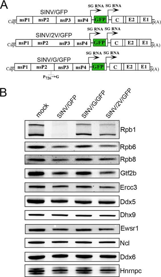
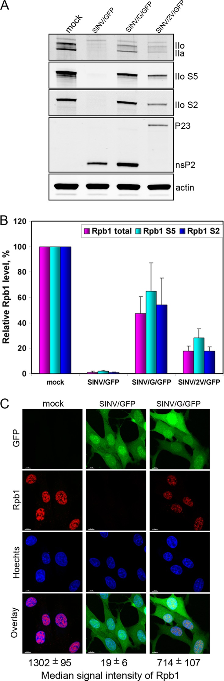
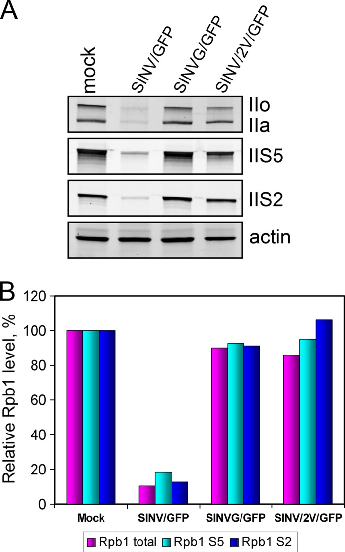
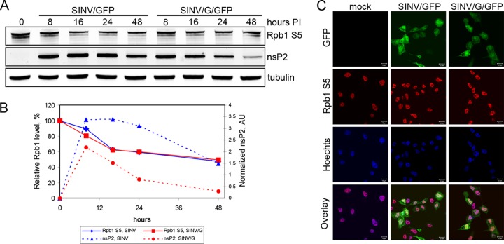
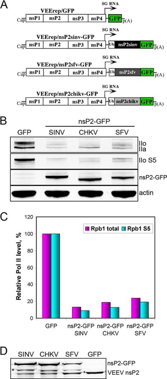
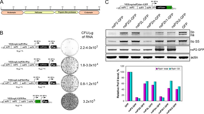
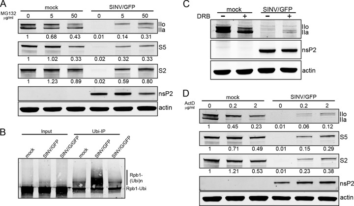
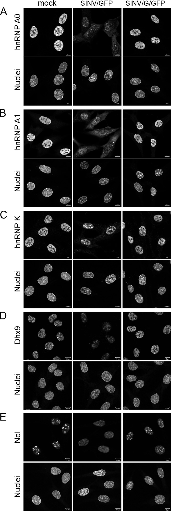
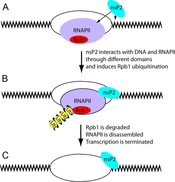
The Old World alphaviruses are emerging human pathogens with an ability to cause widespread epidemics. The latest epidemic of Chikungunya virus, from 2005 to 2007, affected over 40 countries in Africa, Asia, and Europe. The Old World alphaviruses are highly cytopathic and known to evade the cellular antiviral response by inducing global inhibition of transcription in vertebrate cells. This function was shown to be mediated by their nonstructural nsP2 protein; however, the detailed mechanism of this phenomenon has remained unknown. Here, we report that nsP2 proteins of Sindbis, Semliki Forest, and Chikungunya viruses inhibit cellular transcription by inducing rapid degradation of Rpb1, a catalytic subunit of the RNAPII complex. This degradation of Rpb1 is independent of the nsP2-associated protease activity, but, instead, it proceeds through nsP2-mediated Rpb1 ubiquitination. This function of nsP2 depends on the integrity of the helicase and S-adenosylmethionine (SAM)-dependent methyltransferase-like domains, and point mutations in either of these domains abolish Rpb1 degradation. We go on to show that complete degradation of Rpb1 in alphavirus-infected cells occurs within 6 h postinfection, before other previously described virus-induced changes in cell physiology, such as apoptosis, autophagy, and inhibition of STAT1 phosphorylation, are detected. Since Rpb1 is a subunit that catalyzes the polymerase reaction during RNA transcription, degradation of Rpb1 plays an indispensable role in blocking the activation of cellular genes and downregulating cellular antiviral response. This indicates that the nsP2-induced degradation of Rpb1 is a critical mechanism utilized by the Old World alphaviruses to subvert the cellular antiviral response.


Figures

Fig 1
Fig 1
SINV encoding wt but not mutated nsP2 induces degradation of Rpb1. (A) Schematic representation of viral genomes. (B) BHK-21 cells were infected with SINV/GFP or SINV/G/GFP and SINV/2V/GFP mutants at an MOI of 20 PFU/cell. Cellular lysates were prepared at 7 h p.i. and analyzed by immunoblotting using antibodies against different nuclear proteins (see Materials and Methods for details). Abbreviations: Ddx5, DEAD (Asp-Glu-Ala-Asp) box helicase 5; Ddx6, DEAD (Asp-Glu-Ala-Asp) box helicase 6; Dhx9, DEAH (Asp-Glu-Ala-His) box polypeptide 9; Ercc3, excision repair cross-complementing rodent repair deficiency, complementation group 3 (or XPB, xeroderma pigmentosum group B); Ewsr1, Ewing sarcoma breakpoint region 1; Gtf2b, general transcription factor IIB; Nhrnpc, heterogeneous nuclear ribonucleoprotein C; Ncl, nucleolin; Rpb1, polymerase (RNA) II (DNA-directed) polypeptide A (POLR2A); Rpb6, polymerase (RNA) II (DNA-directed) polypeptide F (POLR2F); Rpb8, polymerase (RNA) II (DNA-directed) polypeptide H (POLR2H).
Fig 2
Fig 2
SINV/GFP induces degradation of all forms of Rpb1. (A) BHK-21 cells were infected as described in the legend to Fig. 1. Lysates were prepared at 7 h p.i. and analyzed by immunoblotting using antibodies recognizing different forms of Rpb1: total Rpb1 (IIa and IIo) and Rpb1 phosphorylated at CTD heptate residue serine 5 (IIo S5) or serine 2 (IIo S2). SINV nsP2 was detected by rabbit polyclonal antibodies. (B) Quantitative analysis of immunoblots presented in panel A. Mean values of three experiments and standard deviations (SDs) are presented. (C) Immunofluorescence analysis of total Rpb1 in BHK-21 cells at 7 h p.i. with different SINVs. GFP expression indicates virus replication. Images represent multiple-intensity projections of six optical sections. The median signal intensities of Rpb1-specific nuclear fluorescence were measured for at least 30 randomly selected cells. Bars, 10 μm.
Fig 3
Fig 3
SINV/GFP, but not variants with mutated nsP2, induces Rpb1 degradation in NIH 3T3 cells. (A) NIH 3T3 cells were infected with SINV/GFP or attenuated mutants SINV/G/GFP and SINV/2V/GFP at an MOI of 20 PFU/cell. Lysates were prepared at 7 h p.i. and analyzed by immunoblotting using antibodies recognizing different forms of Rpb1 (see the legend to Fig. 2 for details). (B) Quantitative analysis of immunoblots presented in panel A.
Fig 4
Fig 4
Rpb1 is completely degraded within the first 8 h postinfection with SINV encoding wt nsP2. BHK-21 cells were infected with SINV/GFP (A) or SINV/G/GFP (B) at an MOI of 20 PFU/cell. Cells were harvested at different times postinfection, and levels of Rpb1 and nsP2 were analyzed by immunoblotting with antibodies against different forms of Rpb1 and SINV nsP2. Immunoblots were quantitated as described in Materials and Methods.
Fig 5
Fig 5
SINV infection does not induce Rpb1 degradation in mosquito cells. (A) C7/10 cells were infected with SINV/GFP or SINV/G/GFP. Cells were collected at different times postinfection and analyzed by immunoblotting using antibodies specific to hyperphosphorylated Rpb1 and SINV nsP2. (B) The results of quantitative analysis of immunoblots presented in panel A. (C) Immunofluorescence analysis of total Rpb1 in C7/10 cells at 24 h p.i. GFP expression indicates virus-infected cells. Images represent multiple-intensity projections of six optical sections. Bars, 10 μm.
Fig 6
Fig 6
nsP2 proteins of different OW alphaviruses induce Rpb1 degradation with similar efficiencies. (A) Schematic representation of noncytopathic VEEV replicons encoding GFP fusions of nsP2 proteins derived from different OW alphaviruses. (B) Analysis of Rpb1 levels at 8 h p.i. with the indicated replicons. GFP-expressing replicon VEErep/GFP was used as a control. nsP2-GFP fusion proteins were detected with anti-GFP antibodies. (C) Results of quantitative analysis of immunoblots presented in panel B. (D) Comparative analysis of expression levels of different, heterologous nsP2 proteins and VEEV-specific nsP2 using mouse MAbs recognizing nsP2 proteins of several alphaviruses. The asterisk marks the product of nsP2-GFP degradation.
Fig 7
Fig 7
NTPase, but not protease activity of SINV nsP2, is essential for Rpb1 degradation. (A) Schematic representation of functional nsP2 domains. (B) Comparative analysis of the efficiency of Purr colony formation by VEEV replicons, expressing wt SINV nsP2, its two helicase mutants, and GFP. (C) Analysis of Rpb1 degradation in BHK-21 cells expressing GFP fusions of wt SINV nsP2 or its mutants. Schematic representation of the VEEV-based replicon is shown on the top. (Bottom) Results of quantitative analysis of the immunoblots.
Fig 8
Fig 8
nsP2-induced Rpb1 degradation proceeds through its ubiquitination. (A) SINV/GFP- or mock-infected BHK-21 cells were treated with the indicated concentrations of the proteasome inhibitor MG132 for the entire duration of the infection (6 h). The presence of different forms of Rbp1 was analyzed by immunoblotting. (B) At 2 h p.i., ubiquitinated proteins from mock-, SINV/GFP-, or SINV/G/GFP-infected BHK-21 cells were precipitated with GSK-Dsk2 beads and probed for Rpb1 as described in Materials and Methods. (C and D) Mock- and SINV/GFP-infected BHK-21 cells were treated with 100 μg/ml of DRB (C) or the indicated concentrations of ActD (D) starting from 1 h or 0 h p.i., respectively. Lysates were prepared at 6 h p.i. and analyzed for the presence of Rpb1. Numbers indicate the normalized signal intensities relative to that in untreated, uninfected cells.
Fig 9
Fig 9
Relocalization of several RNA-binding proteins occurs only in the cells infected with the wt nsP2-encoding SINV. Mock-, SINV/GFP-, or SINV/G/GFP-infected BHK-21 cells were immunostained with hnRNP A0 (A)-, hnRNP A1 (B)-, hnRNP K (C)-, Dhx9 (D)- or Ncl (E)-specific Abs at 7 h p.i. Infection of all of the imaged cells was confirmed by detection of GFP, expressed by replicating virus (images are not shown).
Fig 10
Fig 10
Proposed model of nsP2-mediated Rpb1 degradation. (A) nsP2 protein produced by the Old World alphaviruses translocates to the nucleus and binds to DNA. This interaction is mediated by its helicase domain and is most likely not sequence specific. When translocation of RNAPII is stalled at the nsP2-DNA complex, nsP2 interacts with RNAPII through its C domain. (B) This interaction induces polyubiquitination of Rpb1 followed by its degradation and disassembly of the RNAPII complex (C).

References

    1. Agapov EV, et al. 1998. Noncytopathic Sindbis virus RNA vectors for heterologous gene expression. Proc. Natl. Acad. Sci. U. S. A. 95:12989–12994 - PMC - PubMed
    1. Anindya R, Aygun O, Svejstrup JQ. 2007. Damage-induced ubiquitylation of human RNA polymerase II by the ubiquitin ligase Nedd4, but not Cockayne syndrome proteins or BRCA1. Mol. Cell 28:386–397 - PubMed
    1. Atasheva S, Fish A, Fornerod M, Frolova EI. 2010. Venezuelan equine encephalitis virus capsid protein forms a tetrameric complex with CRM1 and importin alpha/beta that obstructs nuclear pore complex function. J. Virol. 84:4158–4171 - PMC - PubMed
    1. Billecocq A, et al. 2004. NSs protein of Rift Valley fever virus blocks interferon production by inhibiting host gene transcription. J. Virol. 78:9798–9806 - PMC - PubMed
    1. Breakwell L, et al. 2007. Semliki Forest virus nonstructural protein 2 is involved in suppression of the type I interferon response. J. Virol. 81:8677–8684 - PMC - PubMed

Publication types