

White matter development in early puberty: a longitudinal volumetric and diffusion tensor imaging twin study
- PMID: 22514599
- PMCID: PMC3326005
- DOI: 10.1371/journal.pone.0032316
White matter development in early puberty: a longitudinal volumetric and diffusion tensor imaging twin study
Abstract
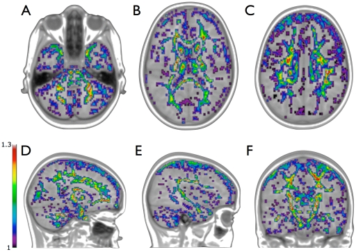
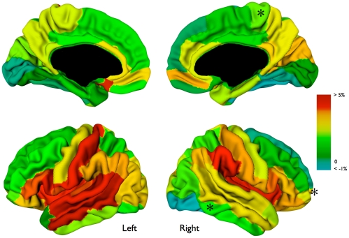
White matter microstructure and volume show synchronous developmental patterns in children. White matter volume increases considerably during development. Fractional anisotropy, a measure for white matter microstructural directionality, also increases with age. Development of white matter volume and development of white matter microstructure seem to go hand in hand. The extent to which the same or different genetic and/or environmental factors drive these two aspects of white matter maturation is currently unknown. We mapped changes in white matter volume, surface area and diffusion parameters in mono- and dizygotic twins who were scanned at age 9 (203 individuals) and again at age 12 (126 individuals). Over the three-year interval, white matter volume (+6.0%) and surface area (+1.7%) increased, fiber bundles expanded (most pronounced in the left arcuate fasciculus and splenium), and fractional anisotropy increased (+3.0%). Genes influenced white matter volume (heritability ~85%), surface area (~85%), and fractional anisotropy (locally 7% to 50%) at both ages. Finally, volumetric white matter growth was negatively correlated with fractional anisotropy increase (r = -0.62) and this relationship was driven by environmental factors. In children who showed the most pronounced white matter growth, fractional anisotropy increased the least and vice-versa. Thus, white matter development in childhood may reflect a process of both expansion and fiber optimization.
Conflict of interest statement
Figures


References
-
- Giedd JN, Blumenthal J, Jeffries NO, Castellanos FX, Liu H, et al. Brain development during childhood and adolescence: a longitudinal MRI study. Nat Neurosci, Vol 2, 1999;861–3 - PubMed
-
- Bartzokis G, Beckson M, Lu PH, Nuechterlein KH, Edwards N, et al. Age-related changes in frontal and temporal lobe volumes in men: a magnetic resonance imaging study. Arch Gen Psychiatry, 58(5), 2001;461–5 - PubMed
-
- van Soelen ILC, Brouwer RM, van Baal GC, Schnack HG, Peper JS, et al. NeuroImage, Epub ahead of print; 2011. Genetic influences on thinning of the cerebral cortex during development. - PubMed
Publication types
MeSH terms
Grants and funding
LinkOut - more resources
Full Text Sources
Other Literature Sources
