. 2012;7(4):e35312.
doi: 10.1371/journal.pone.0035312. Epub 2012 Apr 13.

Endothelial and smooth muscle cells from abdominal aortic aneurysm have increased oxidative stress and telomere attrition

Affiliations

Endothelial and smooth muscle cells from abdominal aortic aneurysm have increased oxidative stress and telomere attrition

Giuseppe Cafueri et al. PLoS One. 2012.

Abstract

Background: Abdominal aortic aneurysm (AAA) is a complex multi-factorial disease with life-threatening complications. AAA is typically asymptomatic and its rupture is associated with high mortality rate. Both environmental and genetic risk factors are involved in AAA pathogenesis. Aim of this study was to investigate telomere length (TL) and oxidative DNA damage in paired blood lymphocytes, aortic endothelial cells (EC), vascular smooth muscle cells (VSMC), and epidermal cells from patients with AAA in comparison with matched controls.

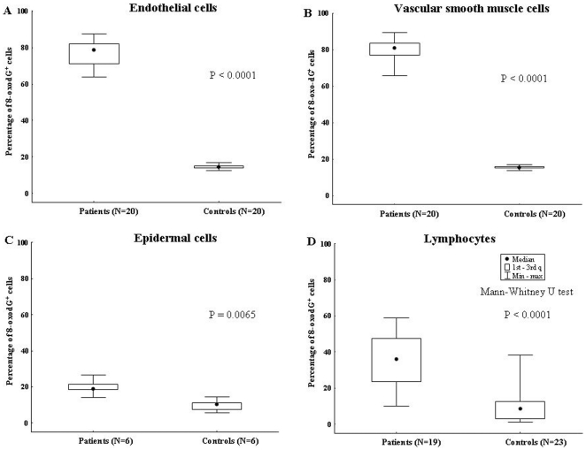
Methods: TL was assessed using a modification of quantitative (Q)-FISH in combination with immunofluorescence for CD31 or α-smooth muscle actin to detect EC and VSMC, respectively. Oxidative DNA damage was investigated by immunofluorescence staining for 7, 8-dihydro-8-oxo-2'-deoxyguanosine (8-oxo-dG).

Results and conclusions: Telomeres were found to be significantly shortened in EC, VSMC, keratinocytes and blood lymphocytes from AAA patients compared to matched controls. 8-oxo-dG immunoreactivity, indicative of oxidative DNA damage, was detected at higher levels in all of the above cell types from AAA patients compared to matched controls. Increased DNA double strand breaks were detected in AAA patients vs controls by nuclear staining for γ-H2AX histone. There was statistically significant inverse correlation between TL and accumulation of oxidative DNA damage in blood lymphocytes from AAA patients. This study shows for the first time that EC and VSMC from AAA have shortened telomeres and oxidative DNA damage. Similar findings were obtained with circulating lymphocytes and keratinocytes, indicating the systemic nature of the disease. Potential translational implications of these findings are discussed.


Conflict of interest statement

Competing Interests: The authors have declared that no competing interests exist.

Figures

Figure 1
Figure 1. Calibration of quantitative FISH analysis.
Telomere fluorescence values (TFU), obtained after hybridization with Cy3 pan-telomeric probe, are converted into kb by external calibration with the L5178Y-S and L5178Y-R murine lymphoma cell lines, MEF murine fibroblast cell line, MCF7 and HeLa human tumor cell lines with known TL of 10.2 Kb, 79.7 Kb, 47 Kb, 4.07 Kb, and 3.44 Kb respectively. Fluorescence intensity (TFU) plotted against the size of TTAGGG repeats sequences (Kb) of the five tumor cell lines. The resulting calibration line was used to transform arbitrary fluorescence intensity units into telomere length in Kb by means of the formula Y = −16.1546+287.2233×X (R = 0.997).
Figure 2
Figure 2. Telomere length of EC and VSMC from patients with AAA measured using Q-FISH and immunofluorescence.
A) Aortic aneurysmatic wall derived EC stained with anti-CD31 mAb (green). The inset shows the nuclei analyzed by Q-FISH. B) EC interphase nuclei hybridized with Cy3-PNA telomeric probe (red signals). C) Aortic aneurysmatic wall derived VSMC stained with anti-α-smooth muscle actin mAb (green). D) VSMC nuclei hybridized with Cy3-PNA telomeric probe (red signals). DAPI was used to label nuclei.
Figure 3
Figure 3. Telomere length of EC, VSMC, blood lymphocytes and epidermal cells from patients with AAA and controls.
A) Telomeres length in EC from 21 AAA patients and in EC from 20 normal aorta. B) Telomeres length in VSMC from 21 AAA patients and in VSMC from 20 normal aorta. C) Telomeres length in epidermal cells from patients with 11 AAA and in these same cells from 6 controls. D) Telomeres length in peripheral blood lymphocytes in 23 AAA patients and in 34 controls.
Figure 4
Figure 4. Telomere length and oxidative DNA damage in lymphocytes from each individual AAA patient and control.
A) Telomere length of peripheral blood lymphocytes from 23 patients with abdominal aortic aneurysms (AAA), and 34 healthy donors. Bars represent the mean. B) Lymphocytes interphase nuclei from an AAA patient hybridized with Cy3-PNA telomeric probe (red signals). C) Oxidative DNA damage in peripheral blood lymphocytes from 19 patients with AAA, and 23 healthy donors. D) A representative immunostaining of anti-8-oxo-dG (green) of blood lymphocytes from an AAA patient. Arrows show nuclei intensively staining for 8-oxo-dG (green). D) Immunostaining of anti-8-oxo-dG (green) of blood lymphocytes from an healthy donor. Arrow shows nucleus with several small positive regions.
Figure 5
Figure 5. Oxidative DNA damage of EC, VSMC, blood lymphocytes and epidermal cells from patients with AAA and controls.
A) The percentage of 8-oxo-dG+ nuclei in EC from 20 AAA patients and in EC from 20 normal aorta. B) The percentage of 8-oxo-dG+ nuclei in VSMC from 20 AAA patients compared to VSMC from 20 normal aorta. C) 8-oxo-dG+ nuclei in epidermal cells from 6 AAA patients and in the same cell type from 6 controls. D) The percentage of 8-oxo-dG+ nuclei in peripheral blood lymphocytes from 19 AAA patients compared to 23 controls.
Figure 6
Figure 6. γH2AX staining patterns observed in EC and VSMC from patients with AAA.
A) Aortic aneurysmatic wall derived EC stained with anti-CD31 (green) and anti-γH2AX (red) mAbs. B) Aortic aneurysmatic wall derived VSMC stained with anti-α-smooth muscle actin (green) and anti-γH2AX (red) mAbs. Focal staining for γH2AX is evident (A, B). Nuclei stained with DAPI (A, B). Magnification, 100× (A, B). C) γH2AX-positive foci were significantly more abundant in EC and VSMC from AAA wall compared to controls (p = 0.006 and p = 0.010 respectively).
Figure 7
Figure 7. Relationship between telomere shortening and DNA damage in blood lymphocytes from AAA patients.
Spearman's correlation test. Linear regression analysis between telomere length and accumulation of ROS-induced oxidative DNA damage, assessed by 8-oxo-dG staining, in blood lymphocytes from 19 AAA patients and from 23 controls. The Spearman's rank correlation coefficient (rS) is  = −0.57.

References

    1. Blackburn EH. Telomere states and cell fates. Nature. 2000;408:53–56. - PubMed
    1. Chan SW, Blackburn EH. New ways not to make ends meet: telomerase, DNA damage proteins and heterochromatin. Oncogene. 2002;21:553–563. - PubMed
    1. Flores I, Benetti R, Blasco MA. Telomerase regulation and stem cell behavior. Curr Opin Cell Biol. 2006;18:254–260. - PubMed
    1. Harley CB, Futcher AB, Greider CW. Telomeres shorten during ageing of human fibroblasts. Nature. 1990;345:458–460. - PubMed
    1. Allsopp RC, Vaziri H, Patterson C, Goldstein S, Younglai EV, et al. Telomere length predicts replicative capacity of human fibroblasts. Proc Natl Acad Sci USA. 1992;89:10114–10118. - PMC - PubMed

Publication types

MeSH terms