. 2012;7(4):e35376.
doi: 10.1371/journal.pone.0035376. Epub 2012 Apr 13.

Structural basis of the chromodomain of Cbx3 bound to methylated peptides from histone h1 and G9a

Affiliations

Structural basis of the chromodomain of Cbx3 bound to methylated peptides from histone h1 and G9a

Jianbin Ruan et al. PLoS One. 2012.

Abstract

Background: HP1 proteins are highly conserved heterochromatin proteins, which have been identified to be structural adapters assembling a variety of macromolecular complexes involved in regulation of gene expression, chromatin remodeling and heterochromatin formation. Much evidence shows that HP1 proteins interact with numerous proteins including methylated histones, histone methyltransferases and so on. Cbx3 is one of the paralogues of HP1 proteins, which has been reported to specifically recognize trimethylated histone H3K9 mark, and a consensus binding motif has been defined for the Cbx3 chromodomain.

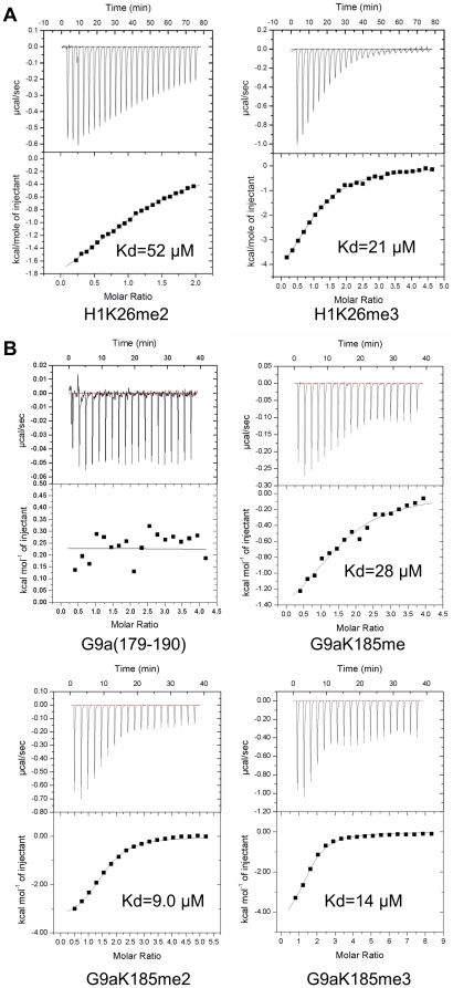
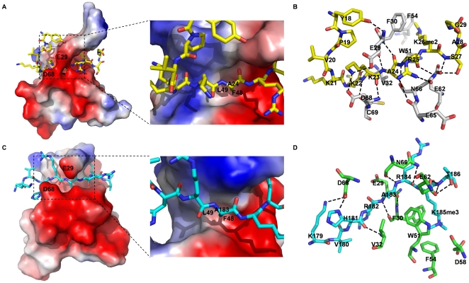
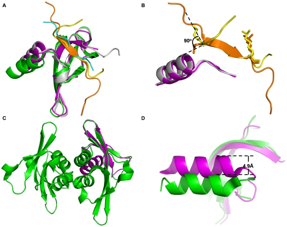
Methodology/principal findings: Here, we found that the Cbx3 chromodomain can bind to H1K26me2 and G9aK185me3 with comparable binding affinities compared to H3K9me3. We also determined the crystal structures of the human Cbx3 chromodomain in complex with dimethylated histone H1K26 and trimethylated G9aK185 peptides, respectively. The complex structures unveil that the Cbx3 chromodomain specifically bind methylated histone H1K26 and G9aK185 through a conserved mechanism.

Conclusions/significance: The Cbx3 chromodomain binds with comparable affinities to all of the methylated H3K9, H1K26 and G9aK185 peptides. It is suggested that Cbx3 may regulate gene expression via recognizing both histones and non-histone proteins.


Conflict of interest statement

Competing Interests: The authors have declared that no competing interests exist.

Figures

Figure 1
Figure 1. Human Cbx3 chromodomain binds to methylated histone H1K26 and G9aK185.
ITC data for Cbx3 chromodomain binding to (A) H1K26 peptides (residues 18–29) and (B) G9aK185 peptides (residues 179–190). Lower panel show fit to a one-site binding model to the binding isotherms.
Figure 2
Figure 2. Structure basis for Cbx3 binding to methylated histone H1K26 and G9aK185 peptide.
(A and C) Electrostatic surface depiction of human Cbx3-histone H1K26me2, and Cbx3-G9aK185me3 complex. Peptide substrates are shown in a stick mode. Surfaces with positive electrostatic potential are blue, and negative potential are red. The side chain of residue H1A24 (G9aA183) inserts into the small hydrophobic pocket formed by Phe48 and Leu49 of human Cbx3. The size of the pocket is only sufficient to accommodate a methyl group but not other residue side chains. (B and D) Binding of histone H1 peptide and G9a peptide in the binding groove of Cbx3 chromodomain, respectively. Hydrogen-bonds are shown as dashed lines. Yellow: histone H1 peptide; Gray: Cbx3 chromodomain in Cbx3-histone H1K26me2 complex. Cyan: G9a peptide; Green: Cbx3 chromodomain in Cbx3-G9aK185me3 complex.
Figure 3
Figure 3. Comparison of three structures of Cbx3 chromodomain binding to methylated histone H3, H1 and G9a peptides.
(A) Superposition of human Cbx3 chromodomain in complex with methylated histone H1 peptide (yellow), histone H3 peptide (orange), G9a peptide (cyan), Cbx3 chromodomains are colored as magenta, gray and green, respectively. (B) Superposition of histone H1 peptide (yellow), histone H3 peptide (orange). (C) Structure of Cbx3-H3K9me3 complex (magenta) was superposed to one protomer of the tetramer of Cbx3-G9aK185me3 complex (green) formed in one asymmetric unit. (D) The α helix (residues 70 to 79) of the chromodomain in the structure of Cbx3-G9aK185me3 complex (green) shifts 4.9 Å away from its counterpart in the structures of Cbx3-H3K9me3 (magenta).

References

    1. Nielsen AL, Oulad-Abdelghani M, Ortiz JA, Remboutsika E, Chambon P, et al. Heterochromatin formation in mammalian cells: interaction between histones and HP1 proteins. Mol Cell. 2001;7:729–739. - PubMed
    1. Canzio D, Chang EY, Shankar S, Kuchenbecker KM, Simon MD, et al. Chromodomain-mediated oligomerization of HP1 suggests a nucleosome-bridging mechanism for heterochromatin assembly. Mol Cell. 2011;41:67–81. - PMC - PubMed
    1. Kim H, Heo K, Choi J, Kim K, An W. Histone variant H3.3 stimulates HSP70 transcription through cooperation with HP1{gamma}. Nucleic Acids Res. 2011;39:8329–8341. - PMC - PubMed
    1. Piacentini L, Pimpinelli S. Positive regulation of euchromatic gene expression by HP1. Fly (Austin) 2010;4:299–301. - PMC - PubMed
    1. Ainsztein AM, Kandels-Lewis SE, Mackay AM, Earnshaw WC. INCENP centromere and spindle targeting: identification of essential conserved motifs and involvement of heterochromatin protein HP1. J Cell Biol. 1998;143:1763–1774. - PMC - PubMed

Publication types