. 2012 Apr 19:9:30.
doi: 10.1186/1742-4690-9-30.

Second-site suppressors of HIV-1 capsid mutations: restoration of intracellular activities without correction of intrinsic capsid stability defects

Affiliations

Second-site suppressors of HIV-1 capsid mutations: restoration of intracellular activities without correction of intrinsic capsid stability defects

Ruifeng Yang et al. Retrovirology. .

Abstract

Background: Disassembly of the viral capsid following penetration into the cytoplasm, or uncoating, is a poorly understood stage of retrovirus infection. Based on previous studies of HIV-1 CA mutants exhibiting altered capsid stability, we concluded that formation of a capsid of optimal intrinsic stability is crucial for HIV-1 infection.

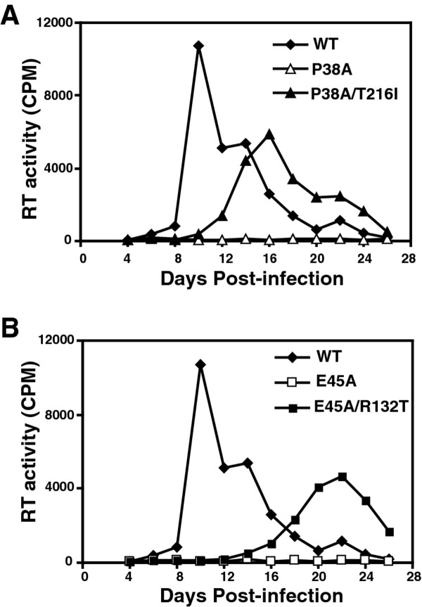
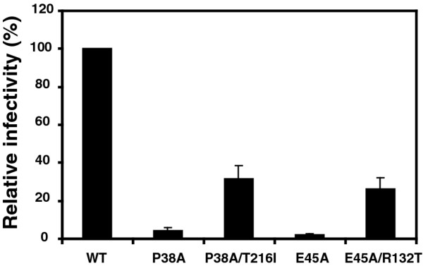
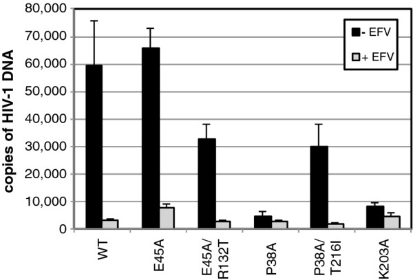
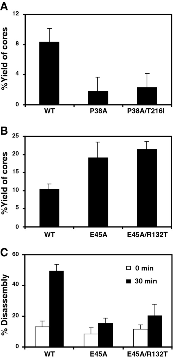
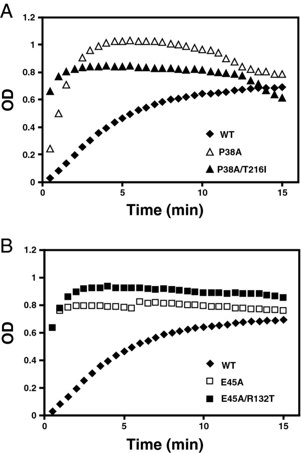
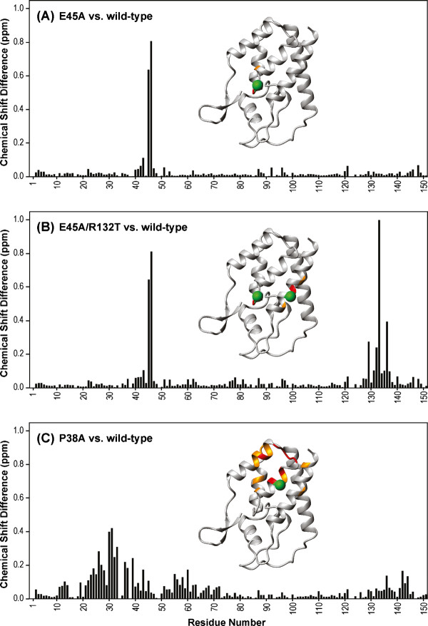
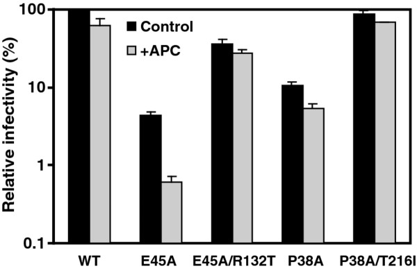
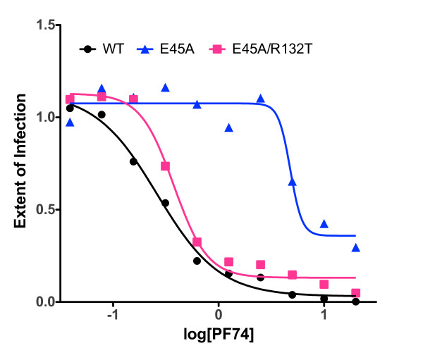
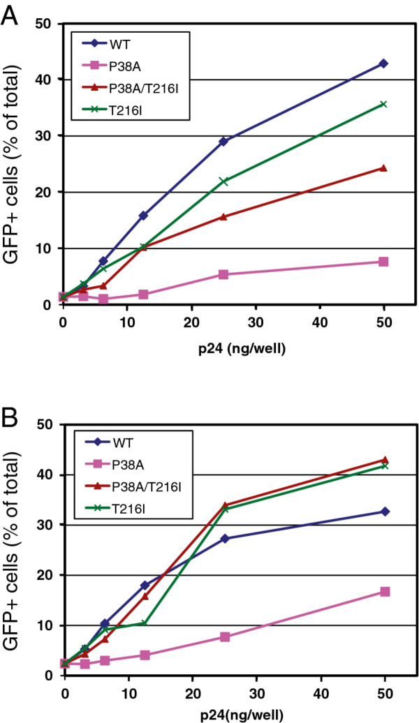
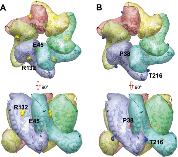
Results: To further examine the connection between HIV-1 capsid stability and infectivity, we isolated second-site suppressors of HIV-1 mutants exhibiting unstable (P38A) or hyperstable (E45A) capsids. We identified the respective suppressor mutations, T216I and R132T, which restored virus replication in a human T cell line and markedly enhanced the fitness of the original mutants as revealed in single-cycle infection assays. Analysis of the corresponding purified N-terminal domain CA proteins by NMR spectroscopy demonstrated that the E45A and R132T mutations induced structural changes that are localized to the regions of the mutations, while the P38A mutation resulted in changes extending to neighboring regions in space. Unexpectedly, neither suppressor mutation corrected the intrinsic viral capsid stability defect associated with the respective original mutation. Nonetheless, the R132T mutation rescued the selective infectivity impairment exhibited by the E45A mutant in aphidicolin-arrested cells, and the double mutant regained sensitivity to the small molecule inhibitor PF74. The T216I mutation rescued the impaired ability of the P38A mutant virus to abrogate restriction by TRIMCyp and TRIM5α.

Conclusions: The second-site suppressor mutations in CA that we have identified rescue virus infection without correcting the intrinsic capsid stability defects associated with the P38A and E45A mutations. The suppressors also restored wild type virus function in several cell-based assays. We propose that while proper HIV-1 uncoating in target cells is dependent on the intrinsic stability of the viral capsid, the effects of stability-altering mutations can be mitigated by additional mutations that affect interactions with host factors in target cells or the consequences of these interactions. The ability of mutations at other CA surfaces to compensate for effects at the NTD-NTD interface further indicates that uncoating in target cells is controlled by multiple intersubunit interfaces in the viral capsid.


Figures

Figure 1
Figure 1
Rescue of P38A (A) and E45A (B) mutants by second-site mutations T216I and R132T, respectively. Replication of mutant and wild-type viruses was analyzed in CEM cells. Samples were collected on the days indicated and analyzed for reverse transcriptase (RT) activity. Shown are the average values obtained from duplicate parallel cultures.
Figure 2
Figure 2
Rescue of P38A and E45A infectivity defects mutations by second-site mutations T216I and R132T. Single-cycle infectivity was assayed in HeLa-P4 target cells. Infectivity was determined as the number of infected cells per ng of p24 in the inoculum, and values are expressed as percentage of wild-type HIV-1 infectivity. Results shown are the mean values of three independent experiments, with error bars representing one standard deviation.
Figure 3
Figure 3
Quantitative analysis of reverse transcription of CA mutants in target cells. HeLa-P4 cells were inoculated with the indicated HIV-1 mutants, and the second-strand transfer viral DNA products accumulated at 8 hours were quantified by PCR. In half of the samples, the reverse transcriptase inhibitor Efavirenz (EFV) was included as a control for plasmid DNA contamination carryover from the transfections used to produce the virus stocks. Error bars represent the standard deviations of triplicate infections, and the results are representative of three independent experiments.
Figure 4
Figure 4
Second-site suppressor mutations do not restore capsid stability in vitro. (A and B) Concentrated virions were subjected to ultracentrifugation through a detergent layer into a sucrose density gradient. Yields of cores were calculated as the percentage of the total CA that was detected in the peak fractions of cores. Results shown are the mean values of three independent experiments, with error bars representing one standard deviation. (C) Disassembly of purified HIV-1 cores in vitro. Diluted HIV-1 cores were incubated at 37°C for the indicated times, followed by separation of free and core-associated CA by ultracentrifugation. Supernatants and pellets were analyzed by p24 ELISA. The extent of disassembly was determined as the percentage of the total CA protein in the reaction detected in the supernatant. Results shown are the average values of two independent experiments with duplicate determinations in each experiment. Error bars represent the spread of values obtained in the two experiments.
Figure 5
Figure 5
In vitro capsid assembly analysis. To initiate assembly, purified recombinant CA proteins were diluted into a buffer, resulting in a final NaCl concentration of 2.25 M. The turbidity of the samples was determined with a spectrophotometer at the indicated times. For reference, the results for the wild type CA protein are shown in both panels A and B. Values shown are the average of two parallel determinations. Shown are data from one representative of 3 independent experiments.
Figure 6
Figure 6
Amide chemical shift differences between the wild type and E45A, E45A/R132T and P38A mutant CA-NTD proteins. Combined 1H, 15 N chemical shift differences between wild-type and mutants E45A (A), E45A/R132T (B) and P38A (C) are plotted versus residue number. Structural mapping of the chemical shift differences onto the X-ray structure of CA-NTD [1AK4] in ribbon representation is provided in the inset. The locations of the mutation sites are marked with large green spheres. Positions of residues whose amide resonances exhibit significant chemical shift differences are color-coded according to the magnitude of the change: red, Δδ > (Δδaverage + 2 × SD); orange, (Δδaverage + 2 × SD) > Δδ > (Δδaverage + 1xSD).
Figure 7
Figure 7
Second-site suppressor mutation R132T relieves the cell-cycle dependence of the E45A mutant in a single-cycle infectivity assay. Control and aphidicolin-arrested HeLa-P4 cells were inoculated with the indicated viruses. Infectivity was determined as described in the legend to Figure 2. Results shown are from one representative of three independent experiments.
Figure 8
Figure 8
Inhibition of wild type, E45A, and E45A/R132T mutant HIV-1 by PF74. Viruses were added to HeLa-P4 target cells at predetermined dilutions in the presence of the indicated concentrations of PF74. Infected cells were identified two days later by X-gal staining and quantified. Results were expressed as the extent of infection normalized by the values obtained with the no-drug controls.
Figure 9
Figure 9
The second-site mutation T216I restores the ability of P38A particles to saturate TRIM5 restrictions in simian cells. Cultures of OMK (A) and FRhK-4 (B) cells were inoculated with the indicated quantities of VSV-G-pseudotyped CA mutant HIV-1 particles and a fixed subsaturating quantity of GFP-encoding pseudotyped HIV-1 particles. Results shown are representative of two independent experiments.
Figure 10
Figure 10
Locations of mutations in the structure of the CA hexamer. (A) Top and side views of a CA hexamer showing E45 (green) and R132 (yellow) side chains from the NMR structure of CA151 (PDB ID: 3H47). (B) Same views, highlighting P38 and T216. P38 (red) in helix 2 is located at the CA NTD-NTD interface and is adjacent to helix 3 in the adjacent subunit. T216 (blue) in helix 11 is near the CA NTD-CTD interface and is close to helix 7 in the nearby subunit.

References

    1. Gamble TR, Vajdos FF, Yoo S, Worthylake DK, Houseweart M, Sundquist WI, Hill CP. Crystal structure of human cyclophilin A bound to the amino-terminal domain of HIV-1 capsid. Cell. 1996;87:1285–1294. doi: 10.1016/S0092-8674(00)81823-1. - DOI - PubMed
    1. Gamble TR, Yoo S, Vajdos FF, von Schwedler UK, Worthylake DK, Wang H, McCutcheon JP, Sundquist WI, Hill CP. Structure of the carboxyl-terminal dimerization domain of the HIV-1 capsid protein. Science. 1997;278:849–853. doi: 10.1126/science.278.5339.849. - DOI - PubMed
    1. Gitti RK, Lee BM, Walker J, Summers MF, Yoo S, Sundquist WI. Structure of the amino-terminal core domain of the HIV-1 capsid protein. Science. 1996;273:231–235. doi: 10.1126/science.273.5272.231. - DOI - PubMed
    1. Li S, Hill CP, Sundquist WI, Finch JT. Image reconstructions of helical assemblies of the HIV-1 CA protein. Nature. 2000;407:409–413. doi: 10.1038/35030177. - DOI - PubMed
    1. Momany C, Kovari LC, Prongay AJ, Keller W, Gitti RK, Lee BM, Gorbalenya AE, Tong L, McClure J, Ehrlich LS. et al.Crystal structure of dimeric HIV-1 capsid protein. Nat Struct Biol. 1996;3:763–770. doi: 10.1038/nsb0996-763. - DOI - PubMed

Publication types

LinkOut - more resources