

Trypanosoma cruzi infection: a review with emphasis on cutaneous manifestations
- PMID: 22515575
- PMCID: PMC3552304
- DOI: 10.1111/j.1365-4632.2011.05380.x
Trypanosoma cruzi infection: a review with emphasis on cutaneous manifestations
Abstract
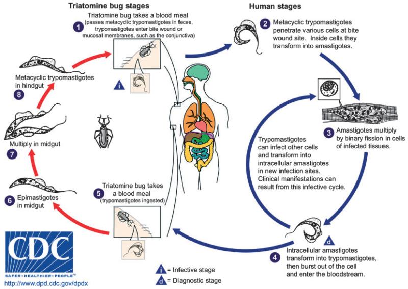
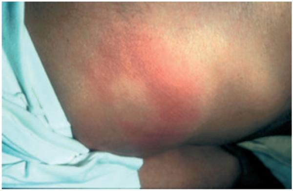
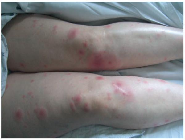
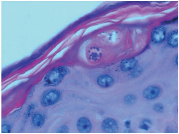
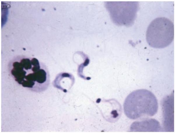
Chagas disease, an infection caused by the protozoan Trypanosoma cruzi and transmitted by the Reduuvid insect vector, remains a major cause of morbidity in Central and South America over a century after its discovery in 1909. Though major advances in preventing the spread of this disease have been made in recent decades, millions of individuals remain chronically infected due to prior exposure to T. cruzi and are at risk for future complications from the disease. Dermatologic manifestations of acute infection may include localized swelling at the site of inoculation (chagoma), conjunctivitis (Romaña's sign), and a generalized morbilliform eruption (schizotrypanides). Reactivation of quiescent infection in immunocompromised hosts due to the acquired immunodeficiency syndrome or organ transplantation can present with fever and skin lesions including panniculitis. The widespread emigration of chronic carriers of T. cruzi to North America, Europe, and Australia makes it imperative that dermatologists worldwide be familiar with this entity to ensure proper diagnosis and treatment.
© 2012 The International Society of Dermatology.
Figures
References
-
- Salvatella R. Current Status of Chagas Disease. Pan American Health Organization; Washington, DC: 2006.
-
- Schmunis GA, Yadon ZE. Chagas disease: a Latin American health problem becoming a world health problem. Acta Trop. 2010;115:14–21. - PubMed
-
- Sartori AM, Sotto MN, Braz LM, et al. Reactivation of Chagas disease manifested by skin lesions in a patient with AIDS. Trans R Soc Trop Med Hyg. 1999;93:631–632. - PubMed
-
- Diaz JH. Recognizing and reducing the risks of Chagas disease (American trypanosomiasis) in travelers. J Travel Med. 2008;15:184–195. - PubMed
-
- Tyler KM, Engman DM. The life cycle of Trypanosoma cruzi revisited. Int J Parasitol. 2001;31:472–481. - PubMed
Publication types
MeSH terms
Grants and funding
LinkOut - more resources
Full Text Sources
Medical
