. 2012 Jul;47(1):126-34.
doi: 10.1016/j.nbd.2012.03.030. Epub 2012 Apr 6.

The location and trafficking routes of the neuronal retromer and its role in amyloid precursor protein transport

Affiliations

The location and trafficking routes of the neuronal retromer and its role in amyloid precursor protein transport

Akhil Bhalla et al. Neurobiol Dis. 2012 Jul.

Abstract

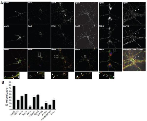
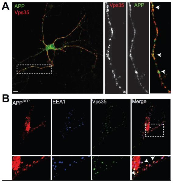
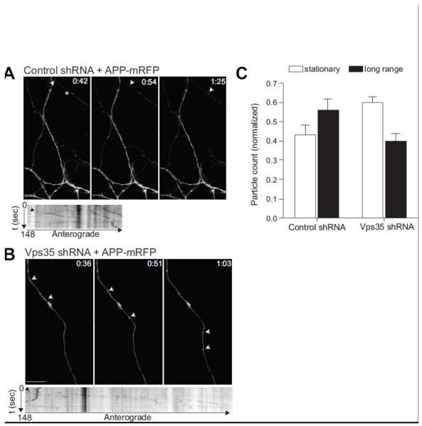
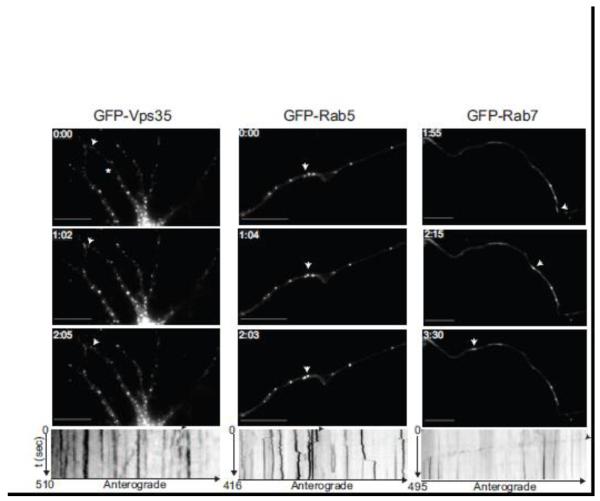
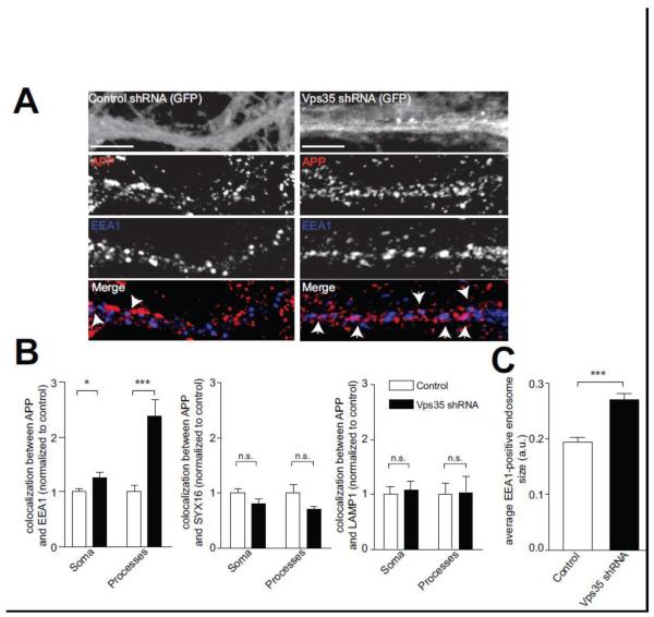
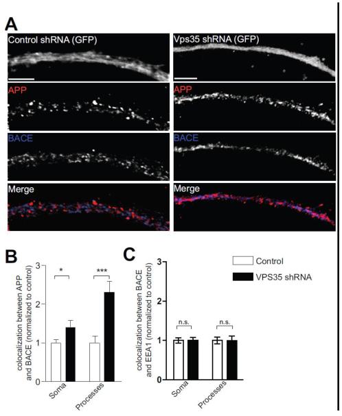
The retromer complex plays an important role in intracellular transport, is highly expressed in the hippocampus, and has been implicated in the trafficking of the amyloid precursor protein (APP). Nevertheless, the trafficking routes of the neuronal retromer and the role it plays in APP transport in neuronal processes remain unknown. Here we use hippocampal neuronal cultures to address these issues. Using fluorescence microscopy, we find that Vps35, the core element of the retromer complex, is in dendrites and axons, is enriched in endosomes and trans-Golgi network, and is found in APP-positive vesicles. Next, to identify the role the neuronal retromer plays in cargo transport, we infected hippocampal neurons with a lentivirus expressing shRNA to silence Vps35. By live fluorescence imaging, Vps35 deficiency was found to reduce the frequency, but not the kinetics, of long-range APP transport within neuronal processes. Supporting the interpretation that retromer promotes long-range transport, Vps35 deficiency led to increased APP in the early endosomes, in processes but not the soma. Finally, Vps35 deficiency was associated with increased levels of Aβ, a cleaved product of APP, increased colocalization of APP with its cleaving enzyme BACE1 in processes, and caused an enlargement of early endosomes. Taken together, our studies clarify the function of the neuronal retromer, and suggest specific mechanisms for how retromer dysfunction observed in Alzheimer's disease affects APP transport and processing.


Figures

Figure 1
Figure 1. Vps35 localizes on endosomes and trans-Golgi Network (TGN) throughout the neuron
A, 14 day old mouse primary hippocampal neurons from postnatal day 0 mice were fixed and stained for endogenous Vps35 (red) and the indicated organelle or synaptic markers (green) using antibodies, and imaged using confocal microscopy (Zeiss LSM 700). Arrows in the inset show regions of colocalization. B, Colocalization index for the indicated stainings. Values denote Means ± SEM (n ≥ 25 fields). Scale bars are 10 μm in all panels.
Figure 2
Figure 2. Vps35 colocalizes with Amyloid Precursor Protein
A, Hippocampal neurons (14 DIV) were fixed and stained for endogenous Vps35 (red) and the Amyloid Precursor Protein (APP) (green). Neurons were imaged as in Fig. 1. Inset shows colocalization between APP and Vps35 occurring also in neuronal processes. B, HeLa cells were transfected with APP-mRFP (red), and stained for endogenous Vps35 (green) and the early endosomal protein EEA1 (blue), 24 hours post transfection. Merge shows extensive colocalization between Vps35 and APP occurring on early endosomes. N=15 fields from at least three independent experiments. Scale bars are 10 μm.
Figure 3
Figure 3. Vps35 deficiency increases Aβ-40 and 42 accumulation
A, Hippocampal neurons (7 DIV) were treated with either control lentivirus or Vps35 shRNA lentivirus, both of which express GFP. Neurons were stained for Vps35 (red) seven days post infection. B, Neurons were treated with lentivirus as in A, and homogenized to obtain a total cell lysate. Western blot analysis shows significant reduction in Vps35 and Vps26 with no significant change in total APP (upper panel) or BACE (lower panel) levels. C, Murine Aβ-40 and 42 levels were measured in the media of either control treated or Vps35 shRNA treated neurons by ELISA (Invitrogen). Values denote Means ± SEM (n=9 from 3 independent experiments). *, p < 0.05. **, p < 0.01 using the Student’s t-test.
Figure 4
Figure 4. APP long-range movement in neuronal processes is affected by Vps35 deficiency
Hippocampal neurons were treated with either control virus (A) or Vps35 shRNA lentivirus (B). Neurons were transfected with APP-mRFP 4 days post viral infection and imaged using an Olympus epi-fluorescence microscope 24-48 hours post transfection. Arrowheads indicate motile APP-mRFP containing organelles. Kymographs (lower panels) were generated using MetaMorph software. The star in A depicts the neuronal process for which the kymograph is shown below. Infected neurons were identified by GFP fluorescence (not shown in the figure). C, A repeated measures analysis of variance was performed including long-range versus stationary particles as the within-group factor and Vps35 kd versus control as the between-group factor. Results showed a significant interaction (F=7.1, p=0.014), such that Vps35 kd caused a relative reduction in long-range particles. N=4 independent experiments. Scale bars are 10 μm.
Figure 5
Figure 5. Vps35 exhibits short-range movement in hippocampal neurons
Hippocampal neurons (9 DIV) were transfected with GFP-Vps35 (left panel), GFP-Rab5 (middle panel) or GFP-Rab7 (right panel), and imaged 24-48 hours post transfection. Time-lapse images were collected using a GFP filter every 5 seconds for the indicated times. Representative time-lapse images show Vps35 (left) positive puncta exhibiting a short-range jitter-like movement similar to Rab5 (middle), whereas Rab7 undergoes long-range movement in neuronal processes. White arrows in the images correspond to the black arrows in the kymographs below, highlighting either the local (for Vps35 and Rab5) or long-range movement (for Rab7). Kymographs were generated from the time-lapse movies using MetaMorph software. The star in Vps35 panel depicts the neuronal process for which the kymograph is shown below. N=3 for Rab5 and Rab7. N>3 for Vps35. Scale bars are 10 μm in all panels.
Figure 6
Figure 6. Vps35 deficiency shifts the localization of APP
A, Hippocampal neurons were treated with control shRNA lentivirus (left panel) or Vps35 shRNA lentivirus (right panel). Neurons were fixed and stained for endogenous APP (red) and the early endosome marker EEA1 (blue). Diffuse GFP fluorescence marks the infected neuronal processes (top most panel) and arrows mark points of colocalization. B, Quantification of the colocalization between APP and EEA1 or APP and LAMP1 in control treated (white bars) or Vps35 shRNA treated (black bars) neurons was determined as done in Fig. 1. Values denote Means ± S.E.M (n>20 fields for Soma and 19 fields for Processes, from 3 independent experiments). Scale bars are 5 μm. C, Quantification of average EEA1 positive endosome size in control and Vps35 shRNA treated neurons was done using ImageJ. Values denote Means ± S.E.M (N=23 from 2 independent experiments). *, p<0.05. ***, p<0.0001 using the Student’s t-test.
Figure 7
Figure 7. Vps35 deficiency increases the colocalization between APP and BACE1
A, Hippocampal neurons were treated with either control shRNA (left panel) or Vps35 shRNA (right panel) as in Fig. 3. Neurons were fixed and stained for endogenous APP (red) and BACE1 (blue). B-C, Colocalization between APP and BACE1 (B), or between BACE1 and EEA1 (C), in control treated (white bars) or Vps35 shRNA treated (black bars) neurons was determined as in Fig. 1C, Values denote Means ± S.E.M (n=17 fields for Soma and 19 fields for Processes, from 3 independent experiments). *, p<0.05. ***, p<0.001 using the Student’s t-test. Scale bars are 5 μm.

References

    1. Andersen OM, Reiche J, Schmidt V, Gotthardt M, Spoelgen R, Behlke J, von Arnim CA, Breiderhoff T, Jansen P, Wu X, Bales KR, Cappai R, Masters CL, Gliemann J, Mufson EJ, Hyman BT, Paul SM, Nykjaer A, Willnow TE. Neuronal sorting protein-related receptor sorLA/LR11 regulates processing of the amyloid precursor protein. Proc Natl Acad Sci U S A. 2005;102:13461–13466. - PMC - PubMed
    1. Arighi CN, Hartnell LM, Aguilar RC, Haft CR, Bonifacino JS. Role of the mammalian retromer in sorting of the cation-independent mannose 6-phosphate receptor. J Cell Biol. 2004;165:123–133. - PMC - PubMed
    1. Belenkaya TY, Wu Y, Tang X, Zhou B, Cheng L, Sharma YV, Yan D, Selva EM, Lin X. The retromer complex influences Wnt secretion by recycling wntless from endosomes to the trans-Golgi network. Dev Cell. 2008;14:120–131. - PubMed
    1. Bonifacino JS, Hurley JH. Retromer. Curr Opin Cell Biol. 2008;20:427–436. - PMC - PubMed
    1. Brunholz S, Sisodia S, Lorenzo A, Deyts C, Kins S, Morfini G. Axonal transport of APP and the spatial regulation of APP cleavage and function in neuronal cells. Exp Brain Res. 2011 - PMC - PubMed

Publication types

MeSH terms

Substances