. 2012 Jul 20;420(4-5):366-83.
doi: 10.1016/j.jmb.2012.04.011. Epub 2012 Apr 16.

Structural and functional characterization of microcin C resistance peptidase MccF from Bacillus anthracis

Affiliations

Structural and functional characterization of microcin C resistance peptidase MccF from Bacillus anthracis

Boguslaw Nocek et al. J Mol Biol. .

Abstract

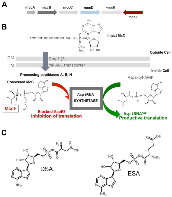
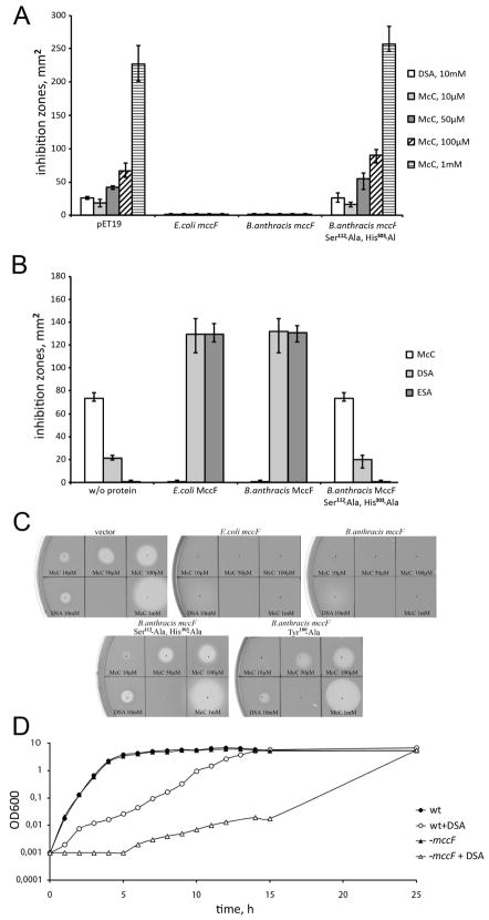
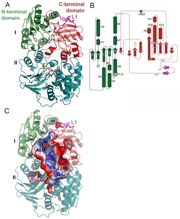
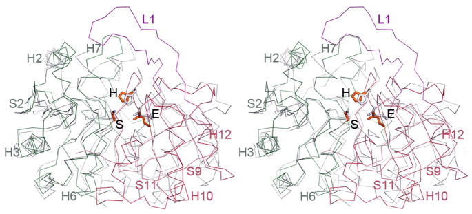
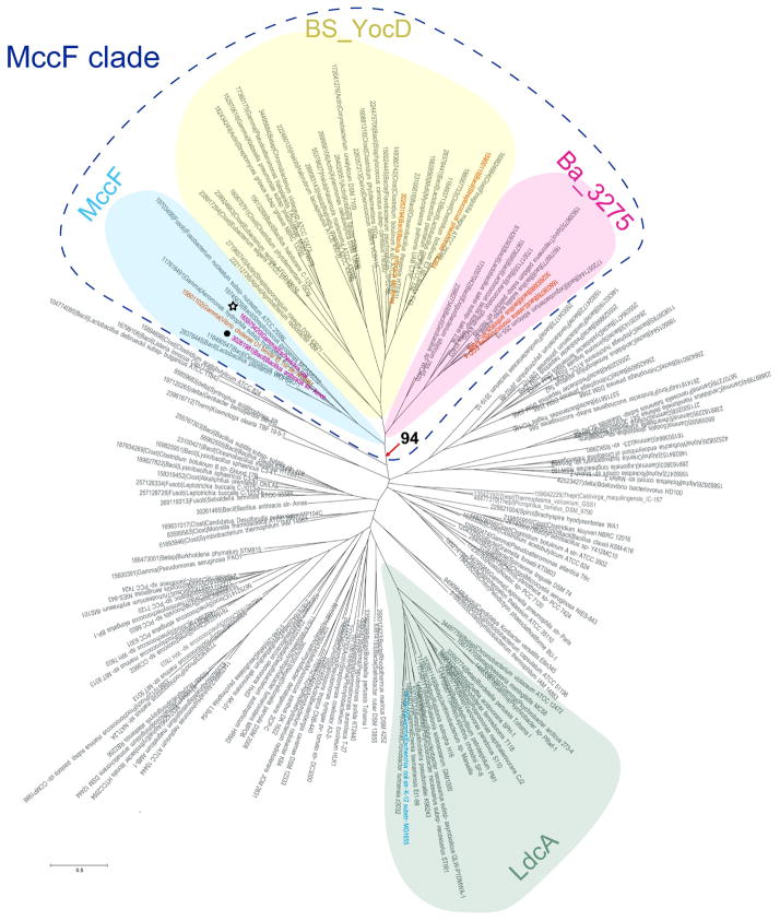
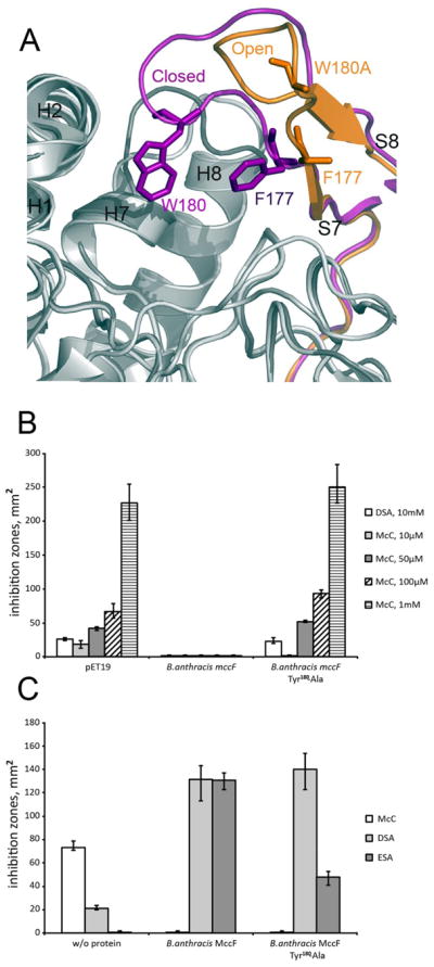
Microcin C (McC) is heptapeptide adenylate antibiotic produced by Escherichia coli strains carrying the mccABCDEF gene cluster encoding enzymes, in addition to the heptapeptide structural gene mccA, necessary for McC biosynthesis and self-immunity of the producing cell. The heptapeptide facilitates McC transport into susceptible cells, where it is processed releasing a non-hydrolyzable aminoacyl adenylate that inhibits an essential aminoacyl-tRNA synthetase. The self-immunity gene mccF encodes a specialized serine peptidase that cleaves an amide bond connecting the peptidyl or aminoacyl moieties of, respectively, intact and processed McC with the nucleotidyl moiety. Most mccF orthologs from organisms other than E. coli are not linked to the McC biosynthesis gene cluster. Here, we show that a protein product of one such gene, MccF from Bacillus anthracis (BaMccF), is able to cleave intact and processed McC, and we present a series of structures of this protein. Structural analysis of apo-BaMccF and its adenosine monophosphate complex reveals specific features of MccF-like peptidases that allow them to interact with substrates containing nucleotidyl moieties. Sequence analyses and phylogenetic reconstructions suggest that several distinct subfamilies form the MccF clade of the large S66 family of bacterial serine peptidases. We show that various representatives of the MccF clade can specifically detoxify non-hydrolyzable aminoacyl adenylates differing in their aminoacyl moieties. We hypothesize that bacterial mccF genes serve as a source of bacterial antibiotic resistance.


Figures

Figure 1
Figure 1
A Organization of the mcc gene cluster from E. coli. Arrows indicate mcc genes and the directions of the arrows indicate the direction of transcription (arrows are not drawn to scale). The mccA–E genes form a single operon and are involved in the synthesis of microcin C (chemical structure shown). Genes whose products contribute to McC immunity are highlighted in red. B. The proposed mechanism of McC transport into the cell, intracellular processing, and inhibition of aspartyl-tRNA synthetase. C. Chemical structure of McC analogs aspartyl sulfamoyl adenylate (DSA) and glutamyl sulfamoyl adenylate (ESA).
Figure 2
Figure 2
A. Overproduction of BaMccF renders cell resistant to microcin C and DSA. Cell lawns were made from cultures producing EcMccF, BaMccF and its double active site mutant – S112A, H303A. Cells transformed with the empty pET19 vector for gene overexpression were used as a negative control. The sizes of growth inhibition zones around 2-μl drops of DSA and McC solutions in indicated concentrations deposited on cell lawns are shown. The error bars show standard deviations of measurements obtained in at least three independent experiments. B. BaMccF inactivates microcin C and its analogs in vitro. Purified recombinant EcMccF, BaMccF and S112A, H303A BaMccF were incubated with microcin C and chemical analogs of its processed form – ESA and DSA. Spots of solutions containing reaction products were placed on E.coli BL21 cell lawn. The method is described in “Experimental Procedures”. Sizes of growth inhibition zones recorded after several hours of cell growth are presented. The error bars show standard deviations of measurements obtained in at least three independent experiments. C. In Vivo sensitivity test of B.cereus ATCC4342 and the mutant on a semisolid agar plate. D. mccF mutant of B.cereus ATCC4342 is hypersensitive to processed microcin C analog. WT and mccF B.cereus ATCC4342 overnight cultures were diluted in fresh LB broth and grown in the presence of 500 mM DSA. No DSA was added to the control cultures. At the indicated time points the OD600 of the culture was measured. Representatives of several independent experiments are shown.
Figure 3
Figure 3
Structure of BaMccF. A. Ribbon diagram showing the overall structure of the MccF dimer, with subunits shown in different colors (subunit I in green, magenta, and red; subunit II in teal). Individual domains of the subunit I are labeled and colored in green and red, while a 27 residues long loop (residues 167–194) connecting them is shown in magenta and labeled L1. The catalytic residues are shown as orange sticks. B. Topology diagrams of MccF. The α-helices and β-strands are shown as cylinders and arrows and the color scheme is maintained as in the figure A. The catalytic triad residues are labeled and their position marked with a star. C. Surface rendering as computed by the program PYMOL showing the active site groove in the dimer of BaMccF. The AMP molecules are shown as spheres. Blue and red represent the positive and negative charge potentials at the + 59 kTe−1 and − 59 kTe−1 scales, respectively. Selected secondary elements are labeled.
Figure 4
Figure 4
Superimposition of members of the S66 serine protease family. A. Ribbon diagram showing a subunit of BaMccF (green, red and magenta) overlaid over a subunit of the dimer of the LdcA from P. aeruginosa (PDB code: 2AUM) (gray), which has been identified as having the closest matching folds. The catalytic triad in both structures (BaMccF S112, E269, H303 correspond to LdcA residues: S115, E217, H285) are positioned similarly. Selected secondary elements of BaMccF are highlighted.
Figure 5
Figure 5
Multiple sequence alignment of MccF proteins from B. anthracis and E. coli versus LdcA from P. aeruginosa and putative LdcA from N. aromaticivorans. Sequence identities are highlighted in red and similarities are shown as red letters. Blue letters mark the catalytic triad residues and the letter L describes the active site loops. The corresponding secondary structure of BaMccF is shown on the top (black).
Figure 6
Figure 6
BaMccF active site with AMP. 2Fo–Fc omit map (light blue) contoured at 1s covering the molecule of AMP. Residues involved in catalysis and binding of the AMP molecule are shown as sticks. Distances in Angstroms are shown as dashed lines. All structural figures were prepared using the program PYMOL.
Figure 7
Figure 7
Phylogenetic tree of S66 family of serine peptidases. The tree was constructed using confidently aligned blocks corresponding to conserved regions of multiple alignments of the S66 family of serine peptidases (174 sequences in total, 188 aligned positions; see Supporting material Fig. S1 for details). Three branches of the MccF clade and LcdA branch are shaded. The bootstrap value for the MccF clade is indicated. The EcMccF(black star) and BaMccF (black dot) sequences are indicated by magenta; sequences corresponding to proteins with determined substrate specificity are indicated by orange. Each terminal node of the tree is labeled by the Genbank Identifier (GI) number, five-letter taxonomy code of an organism and full systematic name of an organism. The taxonomy code is as follows: Gamma – Gammaproteobacteria; Beta – Betaproteobacteria; Alpha - Alphaproteobacteria; delta - Deltaproteobacteria; Bacil – Bacilli; Clost – Clostridia; Deino - Deinococcus-Thermus group; Syner - Synergistetes; Therm - Thermotogae; Acido - Acidobacteria group; Gemma - Gemmatimonadetes; Chroo – Chroococcales; Oscil – Oscillatoriales; Proch - Prochlorales; Nosto -Nostocales; Molli – Mollicutes; Spiro - Spirochaetes; Halob – Halobacteria; Actin – Actinobacteria; Fusob – Fusobacteria; Fibro – Fibrobacteres; Bacte - Bacteroidetes; Negat - Negativicutes; Thepl – Thermoplasmatales; Thepr – Thermoproteales.
Figure 8
Figure 8
A.The active site loop L1. Two conformation of the active site loop L1 observed in the apo form (magenta) and in the W180A mutant (orange) of BaMccF. B. The active site loop L1 W180A mutant shows decreased activity in vivo. Cell lawns were made from cultures producing BaMccF, and the adenosine interacting residue mutant –W180A. Cells transformed with the empty pET19 vector for gene overexpression were used as a negative control. Sizes of growth inhibition zones with around 2-μl drops of DSA and McC solutions in indicated concentrations deposited on cell lawns are shown. The error bars show standard deviations of measurements obtained in at least three independent experiments. C. The active site loop L1 W180A substitution compromise BaMccF activity in vitro. Purified recombinant BaMccF, and W180A BaMccF were incubated with microcin C and chemical analogs of its processed form – ESA and DSA. Spots of solutions containing reaction products were placed on E.coli BL21 cell lawn. The method is described in “Experimental Procedures”. Sizes of growth inhibition zones recorded after several hours of cell growth are presented. The error bars show standard deviations of measurements obtained in at least three independent experiments.

References

    1. Baquero F, Bouanchaud D, Martinez-Perez MC, Fernandez C. Microcin plasmids: a group of extrachromosomal elements coding for low-molecular-weight antibiotics in Escherichia coli. J Bacteriol. 1978;135:342–7. - PMC - PubMed
    1. Severinov K, Semenova E, Kazakov A, Kazakov T, Gelfand MS. Low-molecular-weight post-translationally modified microcins. Mol Microbiol. 2007;65:1380–94. - PubMed
    1. Destoumieux-Garzon D, Peduzzi J, Rebuffat S. Focus on modified microcins: structural features and mechanisms of action. Biochimie. 2002;84:511–9. - PubMed
    1. Guijarro JI, Gonzalez-Pastor JE, Baleux F, San Millan JL, Castilla MA, Rico M, Moreno F, Delepierre M. Chemical structure and translation inhibition studies of the antibiotic microcin C7. J Biol Chem. 1995;270:23520–32. - PubMed
    1. Metlitskaya AZ, Katrukha GS, Shashkov AS, Zaitsev DA, Egorov TA, Khmel IA. Structure of microcin C51, a new antibiotic with a broad spectrum of activity. FEBS Lett. 1995;357:235–8. - PubMed

Publication types

MeSH terms