. 2012 May 4;421(2):361-6.
doi: 10.1016/j.bbrc.2012.04.018. Epub 2012 Apr 9.

Live free or die: stretch-induced apoptosis occurs when adaptive reorientation of annulus fibrosus cells is restricted

Affiliations

Live free or die: stretch-induced apoptosis occurs when adaptive reorientation of annulus fibrosus cells is restricted

Rosalyn D Abbott et al. Biochem Biophys Res Commun. .

Abstract

High matrix strains in the intervertebral disc occur during physiological motions and are amplified around structural defects in the annulus fibrosus (AF). It remains unknown if large matrix strains in the human AF result in localized cell death. This study investigated strain amplitudes and substrate conditions where AF cells were vulnerable to stretch-induced apoptosis. Human degenerated AF cells were subjected to 1 Hz-cyclic tensile strains for 24h on uniformly collagen coated substrates and on substrates with 40 μm stripes of collagen that restricted cellular reorientation. AF cells were capable of responding to stretch (stress fibers and focal adhesions aligned perpendicular to the direction of stretch), but were vulnerable to stretch-induced apoptosis when cytoskeletal reorientation was restricted, as could occur in degenerated states due to fibrosis and crosslink accumulation and at areas where high strains occur (around structural defects, delaminations, and herniations).


Figures

Figure 1
Figure 1. Fabrication of 40 μm collagen stripes to restrict cellular reorientation
A silicon wafer was spin coated with a uniform layer of negative photoresist. UV radiation and a photomask was used to selectively expose the photoresist. Areas that were not exposed to the UV radiation become soluble in developer leaving only the desired pattern. PDMS was then cast against the photoresist master to create a relief of the 3D topography. The PDMS containing the microchannel features was then brought in contact with the silicone elastic substrate. A fitc labeled collagen type I solution submerged the PDMS overnight, confirming the collagen stripe pattern (scale bar= 100 μm).
Figure 2
Figure 2. Cytoskeletal remodeling in response to stretch involved reorientation perpendicular to the direction of stretch (90°) through the redistribution of actin and focal adhesions
The discrete fourier transform (DFT) was used to quantify angular distribution of cells. Representative images of unstretched (A) and 20% stretched (B) cells with the corresponding DFT (C and D, respectively), where the location of peaks in the amplitude corresponds to preferential orientation of cells in that direction. Autocorrelation demonstrated that control images were random (E) while the stretch groups displayed a Gaussian pattern (F). Gaussian curves were fit to the DFT of stretched cells with the mean indicating angle of alignment, and standard deviation the amount of variance within an image. Cellular orientation was clear on imaging of control (G) and stretched (H) cells stained with DAPI (blue), Phalloidin (green), and anti-paxillin (red). Scale bar= 100μm in all images, the stretch direction (B,H) was horizontal, represented by the black arrow, and white arrows (G,H) indicate focal adhesion sites randomly around the control cells and redistributed to the poles of stretched cells perpendicular to stretch.
Figure 3
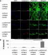
Figure 3. Restricting the adaptive reorientation mechanism, and applying 20% cyclic stretch results in positive staining of cleaved caspase-3, indicating the initiation of an apoptotic pathway
Images of control and stretched cells on either a uniformly collagen coated substrate or restricted to 40 μm collagen stripes stained with DAPI (blue), cleaved caspase-3 (red), and Phalloidin (green) (A). The stretch direction was horizontal and is represented by a white arrow on the images. Scale bars are 50 μm. There was significantly more cleaved caspase-3 staining in the restricted orientation group subjected to 20% cyclic stretch (B, one way ANOVA, p<0.001, Fisher's post hoc test).

References

    1. Bibby SR, Urban JP. Effect of nutrient deprivation on the viability of intervertebral disc cells. Eur Spine J. 2004;13:695–701. - PMC - PubMed
    1. Bibby SR, Jones DA, Ripley RM, Urban JP. Metabolism of the intervertebral disc: effects of low levels of oxygen, glucose, and pH on rates of energy metabolism of bovine nucleus pulposus cells. Spine (Phila Pa 1976) 2005;30:487–496. - PubMed
    1. Roughley PJ. Biology of intervertebral disc aging and degeneration: involvement of the extracellular matrix. Spine (Phila Pa 1976) 2004;29:2691–2699. - PubMed
    1. Adams MA, Roughley PJ. What is intervertebral disc degeneration, and what causes it? Spine (Phila Pa 1976) 2006;31:2151–2161. - PubMed
    1. O'Connell GD, Vresilovic EJ, Elliott DM. Human intervertebral disc internal strain in compression: the effect of disc region, loading position, and degeneration. J Orthop Res. 2011;29:547–555. - PMC - PubMed

Publication types

LinkOut - more resources