. 2012 Jul 1;303(1):C41-51.
doi: 10.1152/ajpcell.00398.2011. Epub 2012 Apr 18.

Increased endogenous H2S generation by CBS, CSE, and 3MST gene therapy improves ex vivo renovascular relaxation in hyperhomocysteinemia

Affiliations

Increased endogenous H2S generation by CBS, CSE, and 3MST gene therapy improves ex vivo renovascular relaxation in hyperhomocysteinemia

Utpal Sen et al. Am J Physiol Cell Physiol. .

Abstract

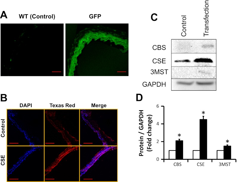
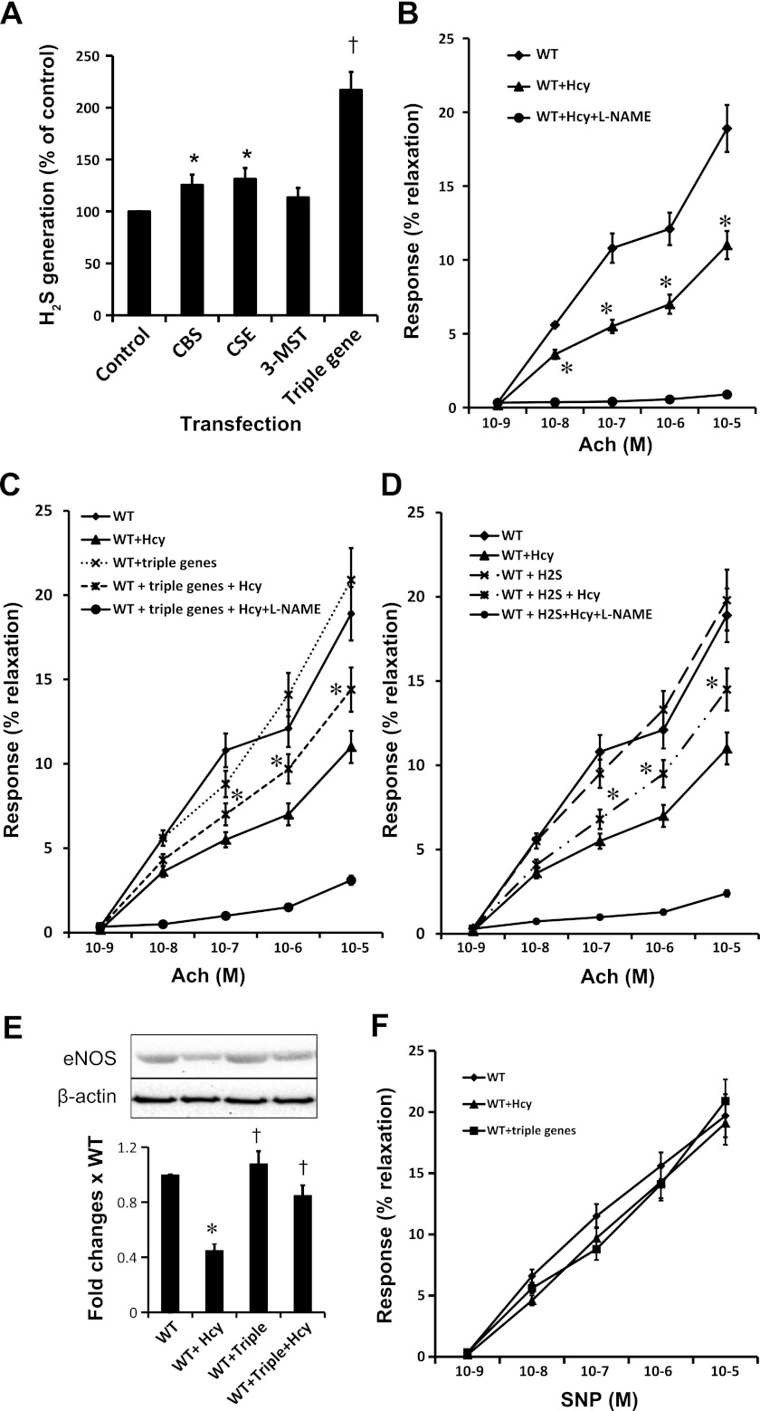
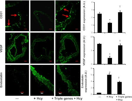
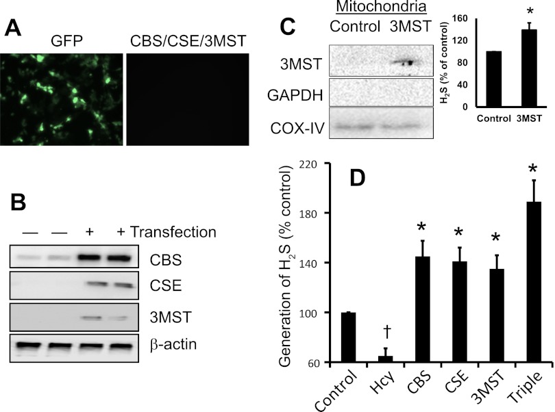
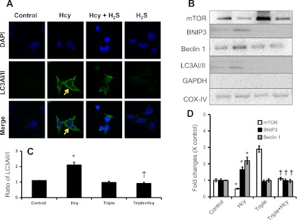
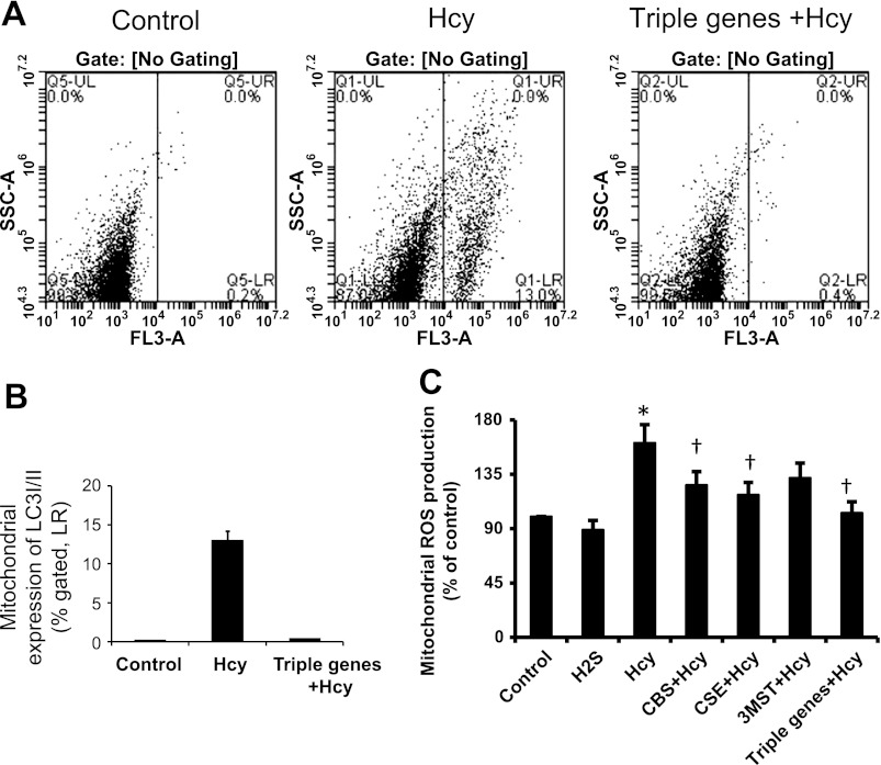
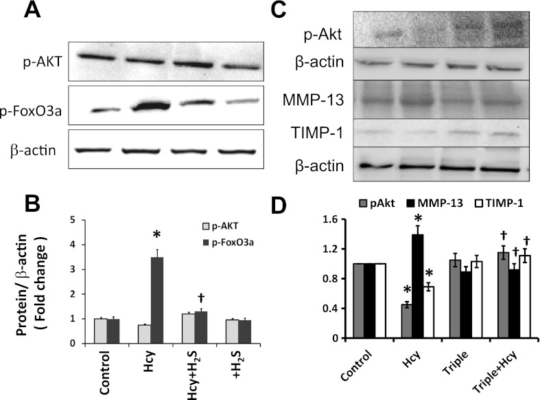
Hydrogen sulfide (H(2)S) has recently been identified as a regulator of various physiological events, including vasodilation, angiogenesis, antiapoptotic, and cellular signaling. Endogenously, H(2)S is produced as a metabolite of homocysteine (Hcy) by cystathionine β-synthase (CBS), cystathionine γ-lyase (CSE), and 3-mercaptopyruvate sulfurtransferase (3MST). Although Hcy is recognized as vascular risk factor at an elevated level [hyperhomocysteinemia (HHcy)] and contributes to vascular injury leading to renovascular dysfunction, the exact mechanism is unclear. The goal of the current study was to investigate whether conversion of Hcy to H(2)S improves renovascular function. Ex vivo renal artery culture with CBS, CSE, and 3MST triple gene therapy generated more H(2)S in the presence of Hcy, and these arteries were more responsive to endothelial-dependent vasodilation compared with nontransfected arteries treated with high Hcy. Cross section of triple gene-delivered renal arteries immunostaining suggested increased expression of CD31 and VEGF and diminished expression of the antiangiogenic factor endostatin. In vitro endothelial cell culture demonstrated increased mitophagy during high levels of Hcy and was mitigated by triple gene delivery. Also, dephosphorylated Akt and phosphorylated FoxO3 in HHcy were reversed by H(2)S or triple gene delivery. Upregulated matrix metalloproteinases-13 and downregulated tissue inhibitor of metalloproteinase-1 in HHcy were normalized by overexpression of triple genes. Together, these results suggest that H(2)S plays a key role in renovasculopathy during HHcy and is mediated through Akt/FoxO3 pathways. We conclude that conversion of Hcy to H(2)S by CBS, CSE, or 3MST triple gene therapy improves renovascular function in HHcy.


Figures

Fig. 1.
Fig. 1.
A: transfection efficacy in arterial explants. Renal arterial explants from wild type (WT; C57BL/6J) were transfected with pcDNA3.1/GFP, pcDNA3.1/CBS, pME18S-CSE-HA, pME18S-3MST, or triple genes of cystathionine β-synthase (CBS), cystathionine γ-lyase (CSE), and 3-mercaptopyruvate sulfurtransferase (3MST) as described in materials and methods and an earlier report (38). After 48 h, GFP-transfected artery was cryosectioned and transfection efficacy was determined by examining pcDNA/GFP fluorescence in fluorescent microscope (scale bar = 50 μm). B: CSE overexpression in vascular smooth muscle cell (VSMC). Since CSE is mainly localized in the VSMC, enhanced expression of CSE in the arterial section after gene delivery was confirmed by immunostaining. Red fluorescence indicated expression of CSE, and blue fluorescence indicated nuclear stain with DAPI (scale bar = 100 μm). C and D: immunoblotting and protein expression levels. Enhanced expression of CBS, CSE, and 3MST protein in the renal arterial explant was further confirmed by immunoblotting following gene transfection (data are means ± SE; n = 3; *P < 0.05 vs. control).
Fig. 2.
Fig. 2.
A: gene delivery increased generation of hydrogen sulfide (H2S) in arterial explants. WT arterial explants after 48 h of gene transfection were homogenized, and tissue capability to generate H2S was determined by our previously adopted method (41) using homocysteine (Hcy) as a substrate. Data represent means ± SE; n = 4–5 independent experiments. *P < 0.05 vs. control; P < 0.01 vs. individual gene transfection. B: Hcy impaired endothelial-dependent vasorelaxation. Renal arterial explants of WT mice were cut into 2-mm pieces and cultured with or without Hcy (75 μM) for 48 h following protocol as described in materials and methods. Arterial rings were mounted in a myobath containing physiological salt solution as described materials and methods. A group of rings that were incubated with Hcy mounted in myobath containing NG-nitro-l-arginine methyl ester (l-NAME; 10 μM), an endothelial nitric oxide synthase (eNOS) inhibitor. Rings were precontracted with phenylephrine (Phe; 10−5M) and later challenged with dose-dependent acetylcholine (Ach) as indicated. Vessels that received l-NAME were virtually unresponsive to Ach, indicating blocked eNOS activity. Data were analyzed by two-way ANOVA and represented as means ± SE; n = 4–5. *P < 0.05 vs. WT at same dose of Ach. C: triple gene therapy improved endothelial-dependent vasorelaxation in HHcy. Renal arterial explants of WT mice were cut into 2-mm pieces, transfected with triple genes and cultured with or without Hcy (75 μM) as indicated for 48 h. Arterial rings of WT and WT + triple genes served as controls for WT + Hcy and WT + triple genes + Hcy, respectively. Rings were mounted between 2 tungsten wires and hung in myobath as described earlier. In another set of experiment, WT arterial rings received l-NAME (10 μM) in the myobath in addition to triple genes and Hcy treatment. Endothelial-dependent vasorelaxation was measured in cumulative Ach doses. Data were analyzed by two-way ANOVA and represent means ± SE; n = 4–5 independent experiments. *P < 0.05 WT + triple genes + Hcy vs. WT + Hcy. D: H2S improved endothelial-dependent vasorelaxation in HHcy. Arterial rings were incubated with Hcy and with or without H2S (30 μM, in the form of NaHS). Endothelial-dependent vasorelaxation was measured as described earlier. Rings that received l-NAME in myobath were unresponsive to Ach, indicating blocked eNOS activity. Data were analyzed by two-way ANOVA and represent means ± SE; n = 4–5 independent experiments. *P < 0.05 WT + H2S + Hcy vs. WT + Hcy. E: expression of eNOS. After myobath study, arterial rings were homogenized in RIPA lysis buffer and eNOS expression was measured by immunoblotting. Data represent means ± SE; n = 4–5. *P < 0.01 vs. WT and P < 0.05 vs. WT + Hcy. F: no changes in endothelial-independent vascular relaxation were observed among the groups. Phe (10−5 M)-precontracted vessels were challenged with dose-dependent (10−9-10−5 M) sodium nitroprusside (SNP), a direct NO donor. No differences were recorded among WT, WT + Hcy, and WT + triple gene vessels, indicating unchanged vascular smooth muscles reactivity among the groups. Data were analyzed by two-way ANOVA and represent means ± SE; n = 4.
Fig. 3.
Fig. 3.
Gene therapy induced CD31 and VEGF and diminished endostatin in renal artery explants in HHcy. CBS, CSE, and 3MST genes were transfected in WT arterial explants, and explants were cultured in matrigel for 48 h in the presence of Hcy (75 μM). Explants were cryosectioned and immunostained with appropriate antibodies secondarily conjugated with FITC. Fluorescence images were taken under confocal microscope (red arrows indicated endothelial lining; scale bar = 50 μm; A.U., arbitrary unit). Bar diagram: data represent means ± SE, n = 7. *P < 0.01 vs. control and P < 0.01 vs. triple genes + Hcy.
Fig. 4.
Fig. 4.
Triple genes expression and H2S generation in endothelial cells. A: mouse aortic endothelial cells (MAECs) were transfected with either CBS, CSE, 3MST, or triple genes. Expression of GFP vector indicating successful transfection. B: after 48 h of transfection, cell were lysed and expression of CBS, CSE, and 3MST protein were measured by Western blot. C: in isolated mitochondria, localized expression of 3MST was confirmed by immunoblotting. Immunoblotting of mitochondria extracted protein with anti-GAPDH antibody indicated isolation of pure mitochondria, which is free from cytosolic fraction of conserved GAPDH protein. Immunoblot was reprobed with cytochrome c oxidase (COX IV) antibody and used as a mitochondrial loading control. The capability of H2S generation by isolated mitochondria from 3MST-transfected cells was measured following protocol as described in materials and methods. Bar diagram showed 3MST-transfected mitochondria generated increased amount of H2S vs. control in presence of Hcy (data are means ± SE; n = 4; *P < 0.05 vs. control). D: cells capability to generate H2S in presence of Hcy was measured as indicated in the method and our previously adopted protocol (37). Representative data from n = 5 independent experiments. Data are means ± SE; n = 4. †P < 0.05 vs. control and *P < 0.05 vs. Hcy.
Fig. 5.
Fig. 5.
Hcy-induced mitophagy was mitigated by H2S and gene therapy. A: for immunostaining, MAECs were cultured for 48 h in the presence or absence of H2S (30 μM, in the form of NaHS) and Hcy (75 μM) as shown. Cells immunostained with microtubule-associated protein light chain 3 (LC3)AI/II antibody secondarily conjugated with FITC (green). Nucleus stained with DAPI (blue). B: Hcy-induced expression of mitophagy markers, mammalian target of rapamycin (mTOR), Beclin 1, BCL2/adenovirus E1B 19-kDa protein-interacting protein 3 (BNIP3), and ratio of LC3AI/II were mitigated by CBS, CSE, and 3MST gene transfection. MAECs were transfected with genes as indicated and treated with or without Hcy (75 μM) for 48 h. Mitochondria were isolated and lysed in RIPA lysis buffer. Equal amounts of protein were analyzed for mitophagy markers as indicated. GAPDH immunoblotting of mitochondria extracted protein indicated isolation of pure mitochondria, which is free from cytosolic GAPDH. Immunoblot was reprobed with COX IV antibody and used as a mitochondrial loading control. Ratio of LC3AII/I (C) and densitometric analyses (D) of mTOR, BNIP3, and Beclin 1 are shown. Data represent means ± SE; n = 4. *P < 0.01 vs. control and †P < 0.01 vs. Hcy.
Fig. 6.
Fig. 6.
Triple gene delivery mitigated mitophagy and mitochondrial reactive oxygen species (ROS) production. A: for flow cytometry, mitochondria were isolated from the treated MAECs as shown, immunostained with LC3AI/II, and analyzed by flow cytometry. Appropriate controls were taken. B: bar diagram showed %mitochondria expressing LC3AI/II marker as an indication of mitophagy. Data represent means ± SE; n = 4/group. *P < 0.05 vs. control and †P < 0.01 vs. Hcy. C: MAECs were transfected either with single gene or triple genes as shown and cultured for 48 h in the presence of Hcy (75 μM). Mitochondria were isolated, and ROS production was detected using DCFDA [5-(and-6)-chloromethyl-2′,7′-dichlorodihydrofluorescein diacetate, acetyl ester] as a substrate. Data represent means ± SE; n = 5 experiments. *P < 0.05 vs. control; P < 0.05 vs. Hcy.
Fig. 7.
Fig. 7.
A and B: Hcy-induced dephosphorylation of Akt and phosphorylation of FoxO3a mitigated by H2S. A: MAECs pretreated with or without H2S (30 μM, in the form of NaHS) were treated with Hcy (75 μM) for 30 min as indicated. A group without any treatment served as control and a group pretreated with H2S served as control for Hcy + H2S. At the end of experiment, cells were lysed and proteins were analyzed by Western blot. B: bar diagram shows densitometric analyses of phospho-protein expression. Data represent means ± SE; n = 4. *P < 0.05 vs. control and †P < 0.05 vs. Hcy treatment. C and D: gene therapy normalized Akt activation and matrix metalloproteinases (MMP)-13/tissue inhibitors of metalloproteinases (TIMP)-1 imbalance. In a separate experiment (C), cells were transfected with CBS, CSE, and 3MST genes and treated with Hcy (75 μM). Expression of phospho-Akt after 30 min of Hcy treatment and MMP-13 and TIMP-1 after 48 h of Hcy treatment was measured by Western blot. D: bar diagram showed densitometric analyses of protein expression. Data represent means ± SE; n = 4. *P < 0.01 vs. control; P < 0.05 vs. Hcy treatment.

References

    1. Ahmed AK, Haylor JL, El Nahas AM, Johnson TS. Localization of matrix metalloproteinases and their inhibitors in experimental progressive kidney scarring. Kidney Int 71: 755–763, 2007 - PubMed
    1. Beard RS, Jr, Bearden SE. Vascular complications of cystathionine β-synthase deficiency: future directions for homocysteine-to-hydrogen sulfide research. Am J Physiol Heart Circ Physiol 300: H13–H26, 2011 - PMC - PubMed
    1. Border WA, Noble NA. TGF-beta in kidney fibrosis: a target for gene therapy. Kidney Int 51: 1388–1396, 1997 - PubMed
    1. Chambers JC, McGregor A, Jean-Marie J, Obeid OA, Kooner JS. Demonstration of rapid onset vascular endothelial dysfunction after hyperhomocysteinemia: an effect reversible with vitamin C therapy. Circulation 99: 1156–1160, 1999 - PubMed
    1. Chang L, Geng B, Yu F, Zhao J, Jiang H, Du J, Tang C. Hydrogen sulfide inhibits myocardial injury induced by homocysteine in rats. Amino Acids 34: 573–585, 2008 - PubMed

Publication types

MeSH terms

Substances