Review
. 2012 Apr 20;12(5):325-38.
doi: 10.1038/nri3198.

Regulation and function of mTOR signalling in T cell fate decisions

Affiliations
Review

Regulation and function of mTOR signalling in T cell fate decisions

Hongbo Chi. Nat Rev Immunol. .

Abstract

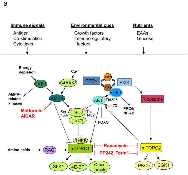
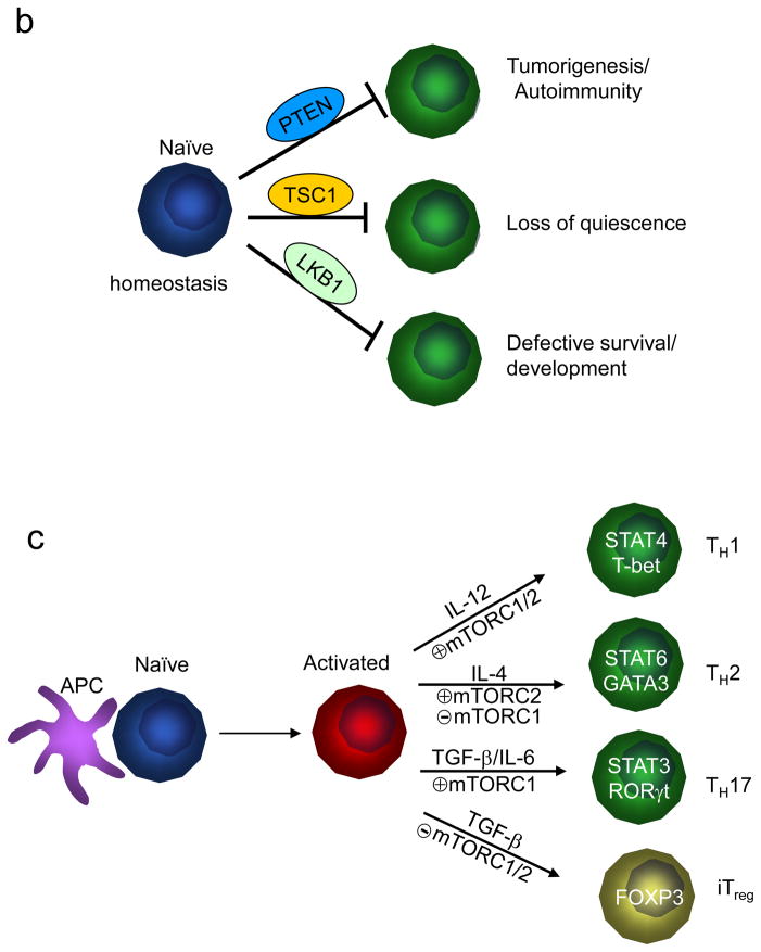
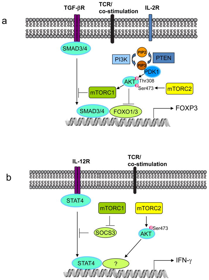
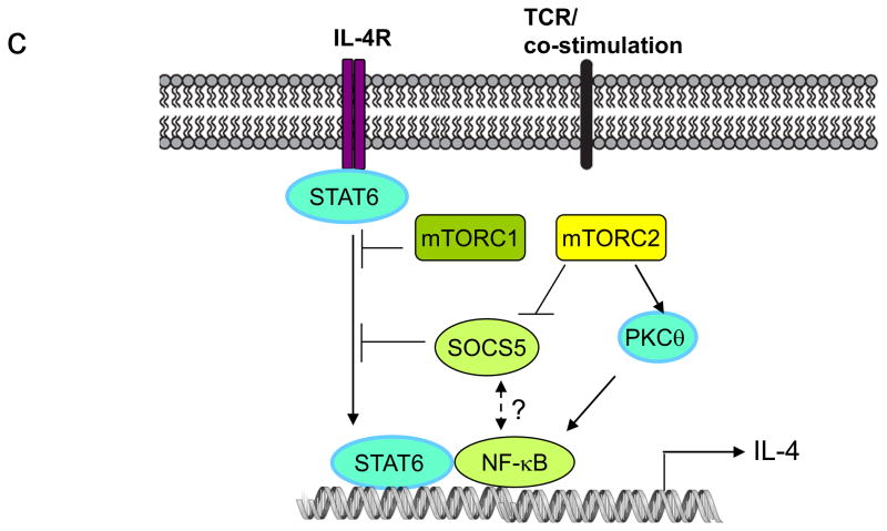
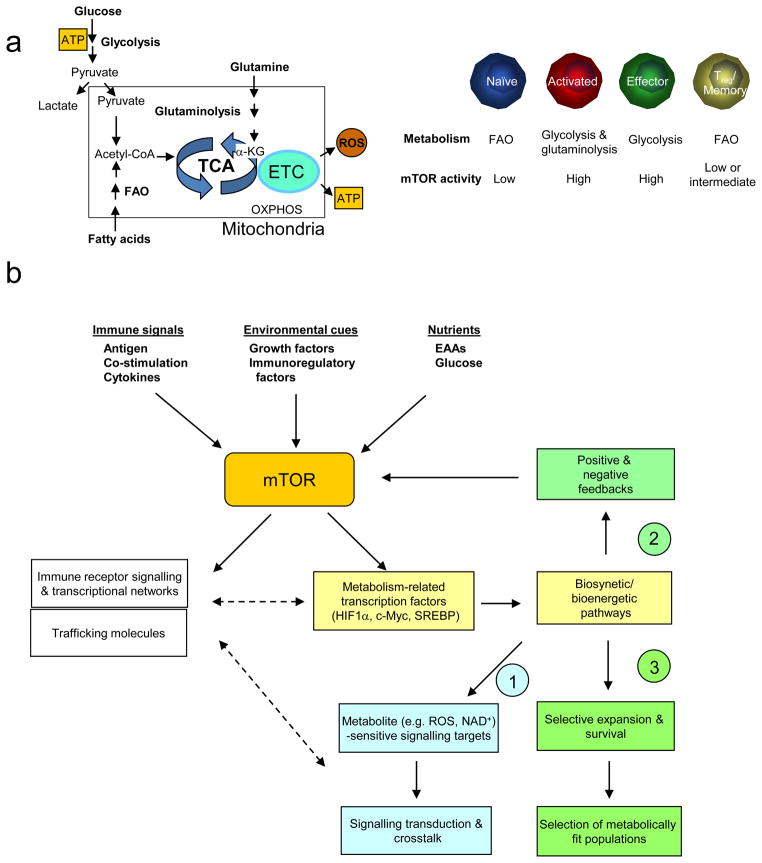
The evolutionarily conserved kinase mTOR (mammalian target of rapamycin) couples cell growth and metabolism to environmental inputs in eukaryotes. T cells depend on mTOR signalling to integrate immune signals and metabolic cues for their proper maintenance and activation. Under steady-state conditions, mTOR is actively controlled by multiple inhibitory mechanisms, and this enforces normal T cell homeostasis. Antigen recognition by naive CD4(+) and CD8(+) T cells triggers mTOR activation, which in turn programmes the differentiation of these cells into functionally distinct lineages. This Review focuses on the signalling mechanisms of mTOR in T cell homeostatic and functional fates, and discusses the therapeutic implications of targeting mTOR in T cells.


Conflict of interest statement

Competing interest statement

The author declares no competing financial interests.

Figures

Figure 1
Figure 1. Regulation and function of mTOR signalling pathways in T cells
(a) Components of mTOR signalling. In T cells, mammalian target of rapamycin (mTOR) can be activated by multiple signals, including the conventionally defined signals 1–3 (antigenic stimulation, co-stimulation and cytokines), growth factors and immunomodulatory factors (such as leptin and sphingosine 1-phosphate (S1P)), and nutrients. The tuberous sclerosis 1 (TSC1)–TSC2 complex integrates signals from phosphoinositide 3-kinase (PI3K)–AKT and liver kinase B1 (LKB1)–AMP-activated protein kinase (AMPK) pathways, and this is mediated by reciprocal regulation of TSC2 activity through AKT-dependent Thr1462 and AMPK-dependent Ser1387 phosphorylation. Upon antigen stimulation, TSC is inactivated by T cell receptor (TCR) signals to mediate mTORC1 activation, but TSC function is maintained in naïve T cells to keep mTOR complex 1 (mTORC1) in check. Additionally, AKT and AMPK can directly modulate mTORC1 functions independently of TSC and RAS homologue enriched in brain (RHEB), and amino acids activate mTORC1 via the RAG family of small GTPases. mTORC1 is best known for its function to promote translation initiation and protein synthesis by directly phosphorylating the substrates S6 kinases (S6Ks) and eIF4E-binding proteins (4E-BPs). Additional mTORC1 targets include the regulatory proteins in cell signalling, metabolism and autophagy. mTORC2 is important for full activation of AKT by inducing Ser473 phosphorylation (thus AKT can be both upstream of mTORC1 and downstream of mTORC2) and for phosphorylation of various protein kinase C (PKC) isoforms including the activation of PKCθ/NF-κB in T cells and serum and glucocorticoid-inducible kinase 1 (SGK1). EAA, essential amino acid; PIP2, phosphatidylinositol-4,5-bisphosphate; PIP3, phosphatidylinositol-3,4,5-triphosphate. (b) Control of T cell homeostasis by active inhibition of mTOR. Under steady state, negative inhibitory molecules for mTOR actively maintain the homeostasis of T cells in the thymus and periphery by preventing them from engaging alternative paths. Although phosphatase and tensin homologue (PTEN), TSC1 and LKB1 have shared capacity to inactivate mTOR, they exert distinct effects to enforce T cell homeostasis. (c) Role of mTORC1 and mTORC2 in functional differentiation of CD4+ T cells. Following antigen stimulation, mTOR signalling promotes differentiation of T helper 1 (TH1), TH2 and TH17 effector cells, and inhibits the induction of regulatory T (TReg) cells.
Figure 2
Figure 2. mTOR-dependent signalling in CD4+ T cell differentiation
(a) Differentiation of induced regulatory T (TReg) cells. Induction of forkhead box P3 (FOXP3) expression depends on the transcription factors SMAD3, SMAD4, FOXO1 and FOXO3. mTOR inhibits induced TReg cell differentiation by antagonizing the function of SMAD3 and SMAD4 downstream of transforming growth factor β (TGF-β) signalling, and by inducing nuclear exclusion of FOXO1 and FOXO3. These two effects are likely mediated by mTOR complex 1 (mTORC1) and mTORC2, respectively. (b) Differentiation of T helper 1 (TH1) cells. mTORC1 inhibits induction of suppressor of cytokine signalling 3 (SOCS3), a crucial negative regulator of signal transducer and activator of transcription 4 (STAT4), to promote interleukin-12 (IL-12) signalling and TH1 cell differentiation. Additionally, mTORC2 is required for activation of AKT that also contributes to interferon γ (IFN-γ) production. (c) Differentiation of TH2 cells. mTORC2 promotes TH2 cell differentiation via two mechanisms: by preventing expression of SOCS5, a negative regulator of IL-4 and STAT6 signalling, and by activating protein kinase C-theta (PKCθ) signalling and nuclear factor-κB (NF-κB)-mediated transcription. By contrast, mTORC1 negatively constrains STAT6 signalling and TH2 cell differentiation.
Figure 3
Figure 3. mTOR in T cell metabolism
(a) Metabolic programmes in T cells (left) and mTOR activity in T cell subsets (right). Only the prominent metabolic programs are presented. FAO, fatty acid oxidation; TCA, tricarboxylic-acid cycle; ETC, electron-transport chain; ROS, reactive oxygen species; OXPHOS, oxidative phosphorylation. (b) Proposed mechanisms of mTOR-mediated metabolic pathways in T cell differentiation. An important upstream signal to activate mTOR is nutrients, in particular essential amino acids (EAAs) whose levels are actively controlled by dendritic cells (DCs). Once activated, mTOR serves as a platform to engage several downstream effector pathways including immune receptor signalling, metabolic programme and migratory activity. mTOR promotes metabolism via activating a gene expression programme consisting of metabolic gene targets of transcription factors hypoxia-inducible factor 1α (HIF1α), c-Myc and SREBP, which in turn impinge upon biosynthetic and bioenergetic pathways. Three potential downstream mechanisms are proposed to explain how these metabolic pathways regulate T cell differentiation, including (1) signal crosstalk; (2) feedback control; (3) selective expansion and survival. Among metabolites with signalling activities, reactive oxygen species (ROS) oxidize the catalytic cysteine residues of phosphatases to cause their inactivation, whereas NAD+ is required for the activity of sirtuin family deacetylases.

References

    1. Zoncu R, Efeyan A, Sabatini DM. mTOR: from growth signal integration to cancer, diabetes and ageing. Nat Rev Mol Cell Biol. 2011;12:21–35. - PMC - PubMed
    1. Thomson AW, Turnquist HR, Raimondi G. Immunoregulatory functions of mTOR inhibition. Nat Rev Immunol. 2009;9:324–337. - PMC - PubMed
    1. Araki K, Ellebedy AH, Ahmed R. TOR in the immune system. Curr Opin Cell Biol. 2011 - PMC - PubMed
    1. Powell JD, Delgoffe GM. The mammalian target of rapamycin: linking T cell differentiation, function, and metabolism. Immunity. 2010;33:301–311. - PMC - PubMed
    1. Iwasaki A, Medzhitov R. Regulation of adaptive immunity by the innate immune system. Science. 2010;327:291–295. - PMC - PubMed

Publication types

Substances