Comparative Study
. 2012 May 15;109(20):E1230-7.
doi: 10.1073/pnas.1119212109. Epub 2012 Apr 19.

Cellular mechanism for selective vertical transmission of an obligate insect symbiont at the bacteriocyte-embryo interface

Affiliations
Comparative Study

Cellular mechanism for selective vertical transmission of an obligate insect symbiont at the bacteriocyte-embryo interface

Ryuichi Koga et al. Proc Natl Acad Sci U S A. .

Abstract

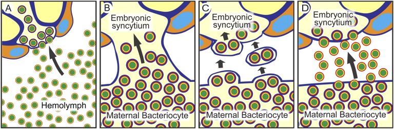
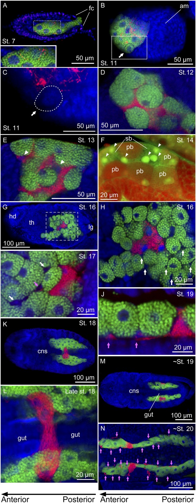
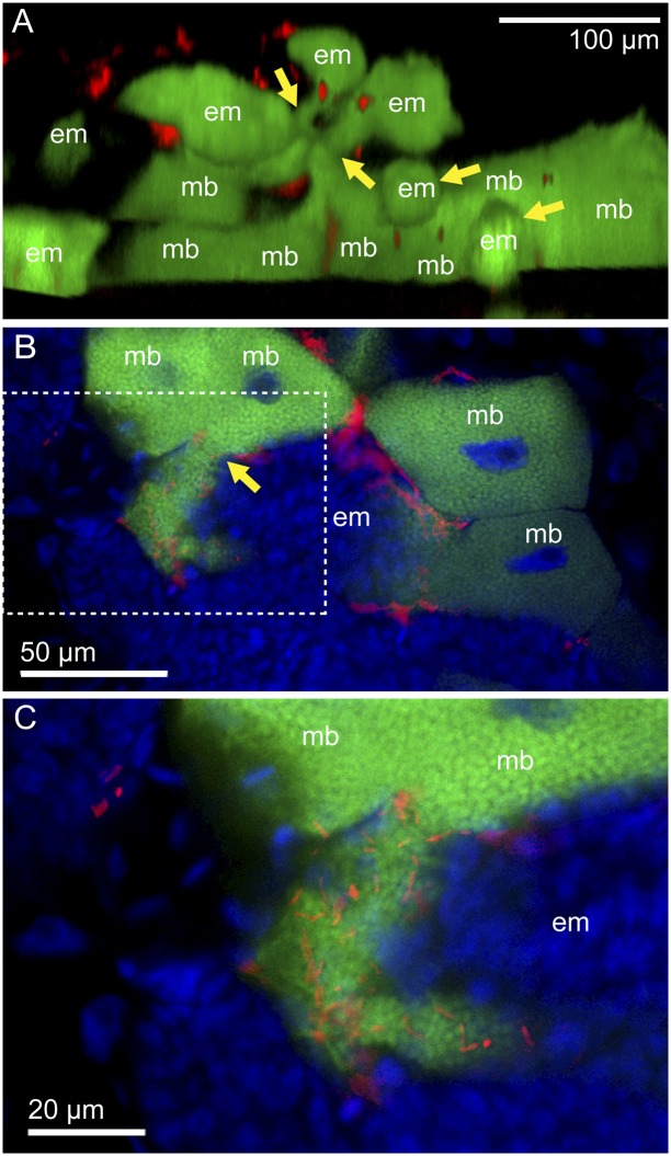
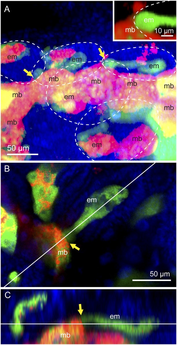
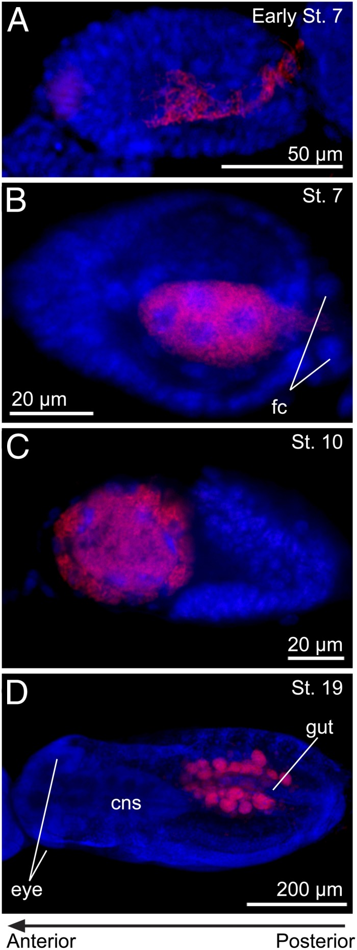
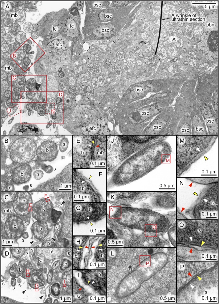
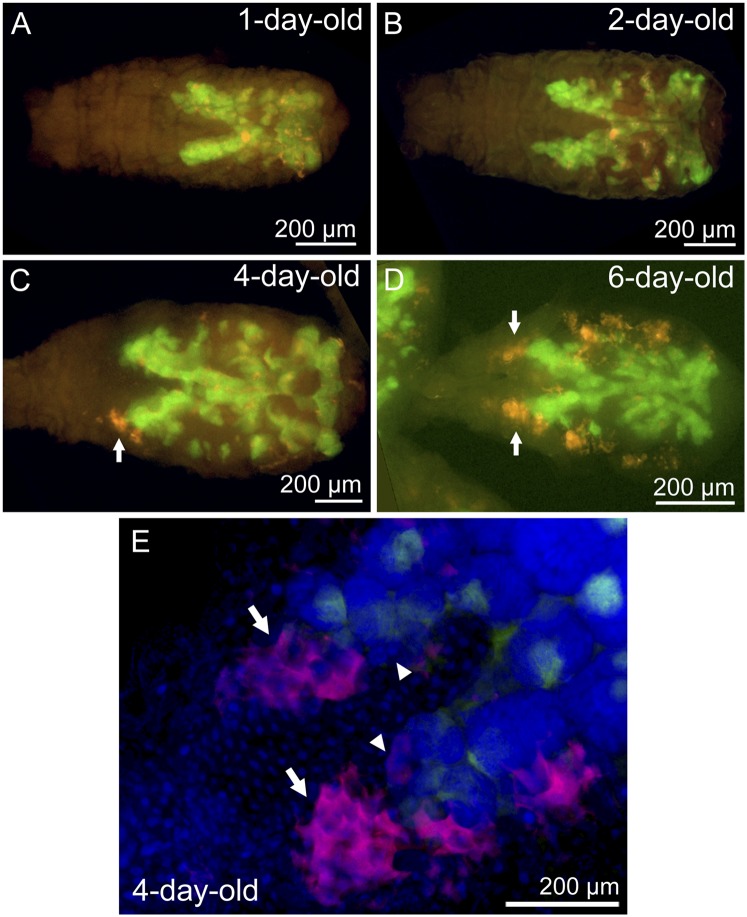
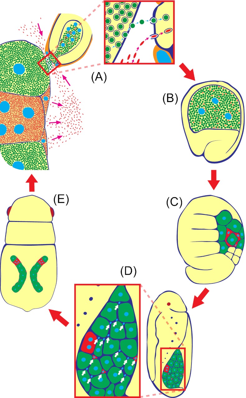
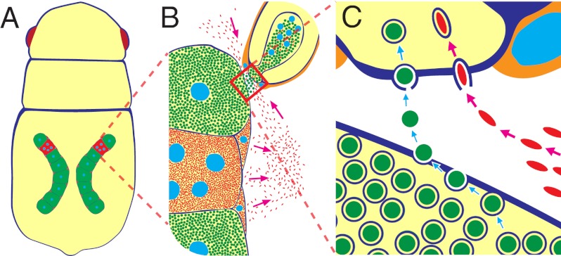
Many insects are associated with obligate symbiotic bacteria, which are localized in specialized cells called bacteriocytes, vertically transmitted through host generations via ovarial passage, and essential for growth and reproduction of their hosts. Although vertical transmission is pivotal for maintenance of such intimate host-symbiont associations, molecular and cellular mechanisms underlying the process are largely unknown. Here we report a cellular mechanism for vertical transmission of the obligate symbiont Buchnera in the pea aphid Acyrthosiphon pisum. In the aphid body, Buchnera cells are transmitted from maternal bacteriocytes to adjacent blastulae at the ovariole tips in a highly coordinated manner. By making use of symbiont-manipulated strains of A. pisum, we demonstrated that the facultative symbiont Serratia is, unlike Buchnera, not transmitted from maternal bacteriocytes to blastulae, suggesting a specific mechanism for Buchnera transmission. EM observations revealed a series of exo-/endocytotic processes operating at the bacteriocyte-blastula interface: Buchnera cells are exocytosed from the maternal bacteriocyte, temporarily released to the extracellular space, and endocytosed by the posterior syncytial cytoplasm of the blastula. These results suggest that the selective Buchnera transmission is likely attributable to Buchnera-specific exocytosis by the maternal bacteriocyte, whereas both Buchnera and Serratia are nonselectively incorporated by the endocytotic activity of the posterior region of the blastula. The sophisticated cellular mechanism for vertical transmission of Buchnera must have evolved to ensure the obligate host-symbiont association, whereas facultative symbionts like Serratia may coopt the endocytotic component of the mechanism for their entry into the host embryos.


Conflict of interest statement

The authors declare no conflict of interest.

Figures

Fig. 1.
Fig. 1.
Hypotheses on the vertical transmission mechanism of the obligate aphid symbiont Buchnera from a maternal bacteriocyte to an early embryo: (A) free symbiont infection, (B) membranous conduit formation, (C) symbiont packet fusion, and (D) exo-/endocytotic transport.
Fig. 2.
Fig. 2.
Infection dynamics of Buchnera and Serratia during embryonic development in the strain IS of A. pisum. In the fluorescent microscopic images, green, red, and blue signals indicate Buchnera cells, Serratia cells, and host insect nuclei, respectively, unless otherwise indicated. Left and right of each panel are anterior and posterior sides, respectively. (A) A stage 7 embryo, to which Buchnera and Serratia are infecting via a posterior passage. Inset: Enlarged image of the dotted rectangle, wherein Serratia cells are clearly seen in addition to Buchnera cells in the central syncytium. (B) A stage 11 embryo in which the symbiont-infected syncytium is cellularizing. Arrow indicates a bacteriocyte that has almost completed the cellularization. (C) An enlarged image of Serratia distribution in the dotted rectangle of B, from which Buchnera signals are removed. Note that no Serratia signals are detected in the completed bacteriocyte (dotted area indicated by an arrow). (D) A late stage 12 embryo in which bacteriocyte formation and symbiont segregation have completed. (E) A stage 13 embryo in which multiple nuclei of the secondary bacteriocyte become evident (arrowheads). (F) A stage 13 embryo stained with green TOTO-3 and red Alexa Fluor 488 phalloidin in which the multiple nuclei (arrowheads) are clearly seen in a large and pleomorphic cytoplasm of the secondary bacteriocyte. (G) A stage 16 embryo in which uninucleate primary bacteriocytes harboring Buchnera and a syncytial secondary bacteriocyte harboring Serratia constitute a large bacteriome. (H) An enlarged image of the bacteriome in the dotted rectangle of G in which a number of small nuclei of sheath cells (white arrows) are seen. (I) The bacteriome of a stage 17 embryo in which Serratia signals appear in the sheath cells (pink arrow). (J) The bacteriome of a stage 19 embryo in which the sheath cells are densely populated by Serratia cells (pink arrows). (K) A stage 18 embryo, whose bacteriome is laterally separating at the region of the secondary bacteriocyte. (L) The bacteriome bridge of a late stage 18 embryo about to be torn apart. (M) A stage 19 embryo in which a pair of syncytial secondary bacteriocytes are established in the bacteriomes. (N) The bacteriomes of a stage 20 embryo. Buchnera is localized in a number of primary bacteriocytes, whereas Serratia is found in a pair of secondary bacteriocytes and many tiny sheath cells (pink arrows). am, amnion; cns, central nervous system; emb, embryo; fc, follicle cell; hd, head; pb, primary bacteriocyte; sb, secondary bacteriocyte; th, thorax.
Fig. 3.
Fig. 3.
Vertical transmission of Buchnera and Serratia to blastulae in the strain IS of A. pisum. (A) Projection image of the bacteriome of a 3-d-old nymph constructed by serial confocal optical sections. Many embryos (em) and maternal bacteriocytes (mb) are seen. Several blastulae are associated with maternal bacteriocytes in the process of symbiont transmission. Yellow arrows indicate the junction sites between blastulae and maternal bacteriocytes. (B) A confocal section of a blastula and maternal bacteriocytes. (C) An enlarged image of the dotted box in B. Green, red, and blue signals indicate Buchnera, Serratia, and host nuclei, respectively. Note that blue signals of host nuclei are not shown in A.
Fig. 4.
Fig. 4.
Selective Buchnera transmission at the bacteriocyte–blastula interface observed in the strain AISTIS of A. pisum. (A) Projection image of the bacteriome of a 3-d-old nymph constructed by serial confocal optical sections. All maternal bacteriocytes (mb) are coinfected with Buchnera (green) and Serratia (red). Several blastulae are associated with maternal bacteriocytes in the process of symbiont transmission (yellow arrows). Note that the embryo contains substantially Buchnera signals only, whereas the maternal bacteriocyte exhibits strong Serratia signals in addition to Buchnera signals. Inset: Highlighted blastula–bacteriocyte junction. Contour of embryos is shown by white dotted circles. (B) A confocal image of another blastula in the process of symbiont transmission. This section corresponds to the plane indicated by a horizontal line in C. (C) A z-axis image constructed from serial confocal sections of the area shown in B. This section corresponds to the plane indicated by a diagonal line in B. Green, red, and blue signals indicate Buchnera, Serratia, and host nuclei, respectively. Yellow arrows show blastula–bacteriocyte junctions.
Fig. 5.
Fig. 5.
Vertical transmission and localization of Serratia in the Buchnera-eliminated strain AISTIS/rif of A. pisum. (A) An early stage 7 blastula in the process of Serratia transmission via the posterior passage. (B) A stage 7 blastula whose central syncytium is filled with Serratia cells. (C) A stage 10 embryo in which the Serratia-infected syncytium is located anteriorly. (D) A stage 19 embryo in which a pair of bacteriome lobes harboring Serratia are located in the abdomen. Red and blue signals indicate Serratia and host nuclei, respectively. cns, central nervous system; fc, follicle cell.
Fig. 6.
Fig. 6.
Transmission EM images of symbiont transmission processes at the bacteriocyte–blastula interface in the IS strain of A. pisum. (A) A low-magnification image of the posterior region of a blastula adjacent to a maternal bacteriocyte, where the symbiont transmission is ongoing. Red rectangles correspond to the locations of the following magnified images. (B) Buchnera cells protruding from the surface of a maternal bacteriocyte at the transmission center. (C) Extracellular Buchnera cells trapped by thin cytoplasmic extensions (black arrowheads) protruding from the posterior pole of the blastula. (D) Buchnera cells endocytosed into the inner syncytial cytoplasm of the blastulae. Serratia cells are also endocytosed into the syncytial cytoplasm. Some of the Buchnera cells look degenerative in the host cytoplasm. (E) Three-layered membrane structure of a Buchnera cell within the maternal bacteriocyte. White, yellow, and red arrowheads indicate bacterial inner membrane, bacterial outer membrane, and host cell membrane, respectively. (F) Two-layered membrane structure of an extracellular Buchnera cell. (G) Two-layered membrane structure of the extracellular side of a Buchnera cell in the process of endocytosis. (H) Three-layered membrane structure of the engulfed side of the same Buchnera cell. (I) Three-layered membrane structure of an endocellular Buchnera cell in the embryonic syncytial cytoplasm. (J) An extracellular Serratia cell in the transmission center. (K) A Serratia cell in the process of endocytosis. (L) A Serratia cell within the syncytial cytoplasm of the blastula. (M) Two-layered membrane structure of an extracellular Serratia cell. (N) Two-layered membrane structure of the extracellular side of a Serratia cell in the process of endocytosis. (O) Three-layered membrane structure of the engulfed side of the same Serratia cell. (P) Three-layered membrane structure of an endocellular Serratia cell in the embryonic syncytial cytoplasm. b, Buchnera cell; bg, presumably degenerating Buchnera cell; bsc, blastula surface cell; efc, enlarged follicle cell; fc, follicle cell; isc, inner syncytial cytoplasm; mb, maternal bacteriocyte; pbn, presumptive bacteriocyte nucleus; s, Serratia cell.
Fig. 7.
Fig. 7.
Localization of Serratia and Buchnera visualized by whole-body in situ hybridization in the IS strain of A. pisum: (A) 1-d-old, first instar nymph; (B) 2-d-old, second instar nymph; (C) 4-d-old, third instar nymph; and (D) 6-d-old, fourth instar nymph. (E) Ovariole tips of a 4-d-old, third instar nymph. Red, green, and blue signals indicate Serratia, Buchnera, and host nuclei, respectively. Note that blue signals of host nuclei are not shown in AD. Arrows show the ovariole tips where Serratia cells aggregate, whereas arrowheads indicate blastulae in which vertical symbiont transmission occurs.
Fig. 8.
Fig. 8.
Infection dynamics of Buchnera and Serratia in the parthenogenetic life cycle of A. pisum. (A) Symbiont transmission from maternal bacteriocyte to blastula. (B) Formation of symbiont-harboring syncytial cytoplasm. (C) Bacteriocyte cellularization and symbiont sorting. (D) Appearance of sheath cells. (E) Establishment of paired bacteriomes. Green, orange, and yellow indicate Buchnera, Serratia, and host embryo, respectively.
Fig. P1.
Fig. P1.
Schematic illustrations of the vertical transmission processes of the obligate symbiont Buchnera (green) and the facultative symbiont Serratia (orange) in the pea aphid A. pisum (yellow). (A) Cellular localization of Buchnera in uninucleate primary bacteriocytes and Serratia in syncytial secondary bacteriocytes and tiny sheath cells. (B) Transmission routes of Buchnera and Serratia to an embryo at the blastula stage. (C) Exo/endocytotic transmission processes of Buchnera and Serratia at the bacteriocyte–blastula interface.

References

    1. Buchner P. Endosymbiosis of Animals with Plant Microorganisms. New York: Interscience; 1965.
    1. Werren JH, Baldo L, Clark ME. Wolbachia: Master manipulators of invertebrate biology. Nat Rev Microbiol. 2008;6:741–751. - PubMed
    1. Oliver KM, Degnan PH, Burke GR, Moran NA. Facultative symbionts in aphids and the horizontal transfer of ecologically important traits. Annu Rev Entomol. 2010;55:247–266. - PubMed
    1. Baumann P. Biology of bacteriocyte-associated endosymbionts of plant sap-sucking insects. Annu Rev Microbiol. 2005;59:155–189. - PubMed
    1. Moran NA, McCutcheon JP, Nakabachi A. Genomics and evolution of heritable bacterial symbionts. Annu Rev Genet. 2008;42:165–190. - PubMed

Publication types

LinkOut - more resources