. 2012 Apr 18;4(130):130ra46.
doi: 10.1126/scitranslmed.3003162.

Dendrimer-based postnatal therapy for neuroinflammation and cerebral palsy in a rabbit model

Affiliations

Dendrimer-based postnatal therapy for neuroinflammation and cerebral palsy in a rabbit model

Sujatha Kannan et al. Sci Transl Med. .

Abstract

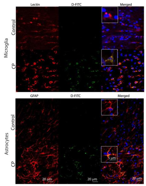
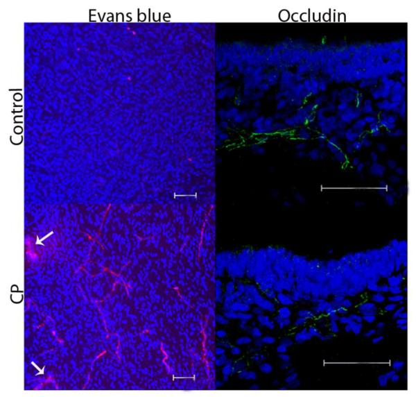
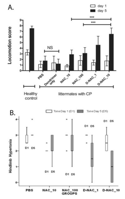
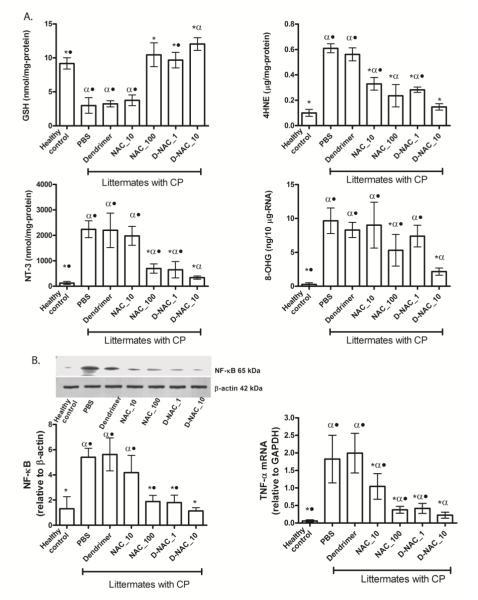
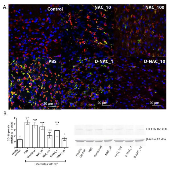
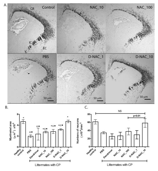
Cerebral palsy (CP) is a chronic childhood disorder with no effective cure. Neuroinflammation, caused by activated microglia and astrocytes, plays a key role in the pathogenesis of CP and disorders such as Alzheimer's disease and multiple sclerosis. Targeting neuroinflammation can be a potent therapeutic strategy. However, delivering drugs across the blood-brain barrier to the target cells for treating diffuse brain injury is a major challenge. We show that systemically administered polyamidoamine dendrimers localize in activated microglia and astrocytes in the brain of newborn rabbits with CP, but not healthy controls. We further demonstrate that dendrimer-based N-acetyl-l-cysteine (NAC) therapy for brain injury suppresses neuroinflammation and leads to a marked improvement in motor function in the CP kits. The well-known and safe clinical profile for NAC, when combined with dendrimer-based targeting, provides opportunities for clinical translation in the treatment of neuroinflammatory disorders in humans. The effectiveness of the dendrimer-NAC treatment, administered in the postnatal period for a prenatal insult, suggests a window of opportunity for treatment of CP in humans after birth.


Figures

Figure 1
Figure 1. Synthesis and characterization of the D-NAC conjugate
Reaction schematic for the synthesis of the dendrimer-NAC (D-NAC) conjugate 1, starting from the free dendrimer and free NAC.
Figure 2
Figure 2. Cellular localization of FITC-labeled dendrimer in the brains of 1 day old newborn healthy (control) and CP rabbits upon intravenous administration
Representative images of the periventricular region from healthy control and endotoxin-exposed newborn rabbits (CP group). Microglia were identified by staining with Tomato lectin (red in microglia panels). Astrocytes were stained with an anti-glial fibrillary acidic protein (GFAP) antibody (red in astrocyte panels). Images were merged to observe co-localization of dendrimer-FITC (D-FITC) with microglia and astrocyte cell types. Nuclei were stained with DAPI (blue). Scale bars, 20 μm. Inset shows microglia and astrocytes at higher magnification (scale bar, 5 μm).
Figure 3
Figure 3. Evaluation of BBB in newborn rabbits with CP
Representative brain sections in the periventricular region from healthy and CP kits on day 1 of life (n=3 per group). The Evans blue-albumin complex is seen as red fluorescence. Arrows point to extravasation of Evans blue-albumin complex into the parenchyma in CP kits. Occludin staining is with Alexa Fluor-488 green.. Nuclei were stained using DAPI (blue). Scale bars, 50 μm.
Figure 4
Figure 4. Motor function and tone in healthy control and CP kits
(A) Locomotion score on day 5 following a single intravenous treatment within 6 h of birth (day 1) of newborn rabbit kits. Data are means and 95% confidence interval (CI) for each treatment group on day 1 and day 5 (n = 6 to 16 endotoxin-exposed kits/treatment group, n=12 healthy kits from 4 litters). See movie S1 (healthy control), movie S2 (PBS treatment), movie S3 (D-NAC_10), movie S4 (dendrimer alone), and movie S5 (NAC_10) for representative locomotor changes between day 1 and day 5. ***P<0.001, NS = not significant. (B) Evaluation of hindlimb tone in rabbit kits on days 1 and 5 of life. Hindlimb tone was assessed in a subset of endotoxin kits following treatment with PBS, NAC (10 and 100 mg/kg), or D-NAC (1 and 10 mg/kg) (n = 5 kits per group) using the modified Ashworth scale: ‘0’ indicates normal tone; ‘1’ indicates slight increase in muscle tone when the limb is moved in extension or flexion; ‘2’ more marked increase in muscle tone through most of the range of movement but affected part is easily moved; ‘3’considerable increase in tone, passive movement is difficult and ‘4’ limb is rigid in flexion or extension. The values are represented as median (indicated by dark horizontal line); inter-quartile range (box); minimum and maximum values (whiskers); and extremes in values or outliers (*).
Figure 5
Figure 5. Oxidative injury and inflammation on day 5 following treatment on day 1
(A) Markers of oxidative injury in periventricular region of the brain. Glutathione (GSH), 4-hydroxynonenal (4HNE), 3-nitrotyrosine (NT-3), and 8-hydroxyguanosine (8-OHG) concentrations were measured in healthy and CP rabbits after treatment (n = 6-7 kits for healthy controls and 4-11 kits for the treatment groups, for each measure). Graphs denote the mean value with 95% CI for each group. *P<0.01 when compared to PBS; #P<0.01 when compared to healthy control; P≤0.01 when compared to D-NAC_10. (B) NF-κB levels and TNF-α mRNA levels in the periventricular region of the brain. Western blot of NF-κB p65 expression was quantified and normalized to β-actin. TNF-α mRNA was quantified and normalized to GAPDH expression. Graphs denote the mean value with 95% CI for each group. *P<0.01 when compared to PBS; #P<0.01 when compared to control; P ≤ 0.05 when compared to D-NAC_10.
Figure 6
Figure 6. Microglial response in the periventricular white matter region of rabbit kits on day 5 of life
(A) To detect a pro-inflammatory microglial phenotype, brain sections were stained using anti-CD11b (green). Tomato lectin (red) counter-stain was used for microglial morphology. Merged areas appear yellow. Scale bar, 20 μm. (B) Western blot for CD11b and its quantification, with CD11b expression normalized to β-actin. Data are means with 95% CI for each group *P<0.05 when compared to PBS; αP<0.01 when compared to healthy control; •P<0.01 when compared to D-NAC_10.
Figure 7
Figure 7. Effect of therapy on myelination and neuronal injury
(A) Myelination in rabbit kits on day 5 of life. Representative brain sections stained for MBP. CR, corona radiata; EC, external capsule; IC, internal capsule. (B) Quantification of myelination. Data are means with 95% CI of the myelinated area per hemisphere. *P<0.05 when compared to PBS; αP<0.01 when compared to control; P<0.01 when compared to D-NAC_10. (C) Neuronal cell count in the caudate region of basal ganglia in the brains of newborn rabbits. Mature neurons were identified by MAP2 staining in the caudate region of the basal ganglia. NS, not significant.

Comment in

References

    1. Rosenbaum P, Paneth N, Leviton A, Goldstein M, Bax M, Damiano D, Dan B, Jacobsson B. A report: the definition and classification of cerebral palsy April 2006. Dev. Med. Child Neurol. 2007;49:s109, 3–7. - PubMed
    1. Kirby RS, Wingate MS, Van Naarden Braun K, Doernberg NS, Arneson CL, Benedict RE, Mulvihill B, Durkin MS, Fitzgerald RT, Maenner MJ, Patz JA, Yeargin-Allsopp M. Prevalence and functioning of children with cerebral palsy in four areas of the United States in 2006: a report from the Autism and Developmental Disabilities Monitoring Network. M. Res Dev Disabil. 2011;32:462–9. - PubMed
    1. Honeycutt A, Dunlap L, Chen H, Homsi G, Grosse S, Schendel D. Economic Costs Associated with Mental Retardation, Cerebral Palsy, Hearing Loss, and Vision Impairment - United States. Morb. Mortal Wkly. Rep. 2004;53:57–59. - PubMed
    1. Leviton A, Gilles F. Maternal urinary tract infections and fetal leukoencephalopathy. N. Engl. J. Med. 1979;301:661. - PubMed
    1. Yoon BH, Romero R, Park JS, Kim CJ, Kim SH, Choi JH, Han TR. Fetal exposure to an intra-amniotic inflammation and the development of cerebral palsy at the age of three years. Am. J. Obstet. Gynecol. 2000;182:675–681. - PubMed

Publication types