. 2012 Jun 14;119(24):e172-80.
doi: 10.1182/blood-2012-03-414557. Epub 2012 Apr 18.

A switch in infected erythrocyte deformability at the maturation and blood circulation of Plasmodium falciparum transmission stages

Affiliations

A switch in infected erythrocyte deformability at the maturation and blood circulation of Plasmodium falciparum transmission stages

Marta Tibúrcio et al. Blood. .

Abstract

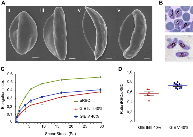
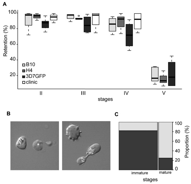
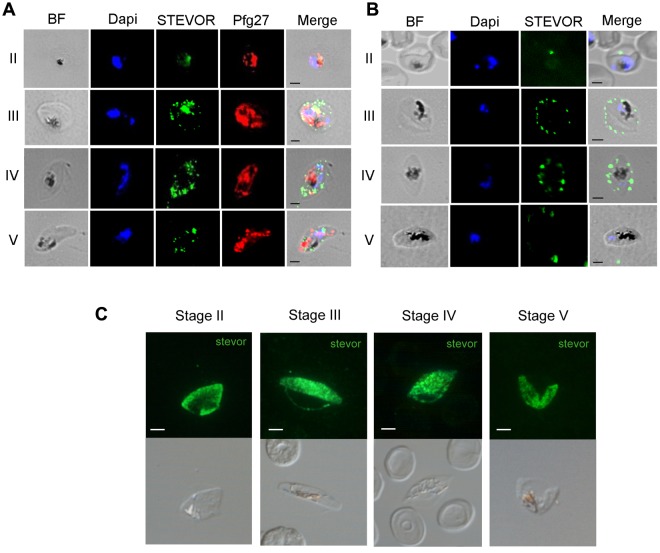
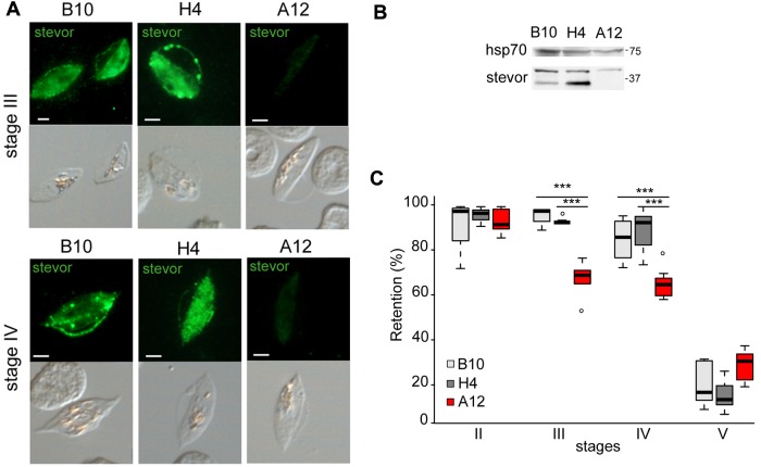
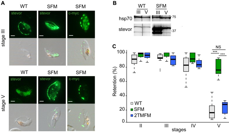
Achievement of malaria elimination requires development of novel strategies interfering with parasite transmission, including targeting the parasite sexual stages (gametocytes). The formation of Plasmodium falciparum gametocytes in the human host takes several days during which immature gametocyte-infected erythrocytes (GIEs) sequester in host tissues. Only mature stage GIEs circulate in the peripheral blood, available to uptake by the Anopheles vector. Mechanisms underlying GIE sequestration and release in circulation are virtually unknown. We show here that mature GIEs are more deformable than immature stages using ektacytometry and microsphiltration methods, and that a switch in cellular deformability in the transition from immature to mature gametocytes is accompanied by the deassociation of parasite-derived STEVOR proteins from the infected erythrocyte membrane. We hypothesize that mechanical retention contributes to sequestration of immature GIEs and that regained deformability of mature gametocytes is associated with their release in the bloodstream and ability to circulate. These processes are proposed to play a key role in P falciparum gametocyte development in the host and to represent novel and unconventional targets for interfering with parasite transmission.


Figures

Figure 1
Figure 1
Scanning electron microscopy and ektacytometry analysis of immature and mature P falciparum GIEs. (A) Scanning electron microscopy images of Plasmodium falciparum GIEs (B10 clone) from stage II to stage V of maturation. Bars represent 1 μm. (B) Giemsa staining images of stage II/III (top panel) and stage V (bottom panel) GIE samples used in ektacytometry analysis. (C) Response to increasing shear stress of erythrocytes infected by P falciparum stage II/III (red line) and stage V (blue line) gametocytes (40% parasitemia) and of uninfected erythrocytes (green line). Error bars represent SE. (D) Ratios of EIs of infected versus uninfected erythrocytes calculated from the 3- to 9.49-Pa range of shear stresses in the ektacytometry analysis (C) showing a statistically significant difference between immature and mature GIEs (Mann-Whitney rank-sum test, P = .0004).
Figure 2
Figure 2
Retention in microsphilters of immature and mature GIEs. (A) Retention in microsphilters of stages II, III, IV, and V GIE from different P falciparum clonal lines (B10, H4, and 3D7GFP) in culture or directly collected from the blood of a hyposplenic patient treated for a malaria attack (clinic). Immature GIEs (stages II-IV) are retained by the microsphilters while mature GIEs (stage V) flow through. (B) Differential interference contrast images of paraformaldehyde-fixed GIEs as they flow through the microsphilters. Immature stages (left panel) keep a convex oval or round shape, unlike uninfected erythrocytes (white star), whereas a majority of mature GIEs are twisted and dumbbell-shaped (right panel). (C) Graphical representation for the proportion of GIEs showing a regular (dark gray) or twisted (light gray) shape in a population of immature (n = 100) and mature (n = 36) GIEs (χ2 test, P = 1 × 10⁻⁹).
Figure 3
Figure 3
Immunofluorescence analysis of STEVOR protein expression in GIEs. (A) Analysis of STEVOR protein expression in fixed stages II to V GIE preparations. GIEs were acetone-fixed and costained with mouse anti-S2 (green) and rabbit anti-Pfg27 (red) followed by anti–mouse Alexa-488– and anti–rabbit Alexa-594–conjugated IgG. Parasite nuclei were stained with DAPI (blue). Bright-field (BF) and merge images are shown. Bars represent 2 μm. (B) Analysis of live GIEs immunostained with rabbit anti-S2 (green) specifically recognizing native STEVOR on the surface of stages III and IV GIEs. Bound antibody was detected with anti–rabbit Alexa-488–conjugated IgG. BF, nuclear staining (DAPI, blue), and merge images are shown. (C) Immunofluorescence analysis of STEVOR expression in fixed stages II to V-GIEs. GIEs were methanol-fixed and stained with a pool of mouse polyclonal antibodies directed against STEVOR proteins followed by anti–mouse Alexa-488–conjugated IgG (green). Bars represent 2 μm.
Figure 4
Figure 4
Decrease in STEVOR expression is associated with an increase in deformability of immature GIEs. (A) Immunofluorescence analysis of STEVOR expression in stage III and stage IV GIEs from the B10, H4, and A12 clones. GIEs were stained with a pool of mouse polyclonal antibodies directed against STEVOR proteins followed by anti–mouse Alexa-488–conjugated IgG. Signal intensity was analyzed by ImageJ Version 6 on at least 30 pictures taken under identical exposition conditions for each clone. Bars represent 2 μm. (B) Western blot analysis of STEVOR expression in stage III and stage V GIEs from the wild-type and the SFM parasites. Immunoblots were probed with a pool of mouse polyclonal antibodies directed against STEVOR proteins and with a mAb directed against HSP70. (C) Retention in microsphilters of stages II, III, IV, and V GIEs from the B10 (light gray), H4 (dark gray), and A12 (red) clones. ***Highly significant differences in retention rates (P < .01). Outliers are shown as open circles.
Figure 5
Figure 5
Stevor overexpression is associated with a decrease in deformability of mature GIEs. (A) Immunofluorescence analysis of STEVOR expression in stage III and stage V GIEs from the wild-type (WT) and the SFM parasites. GIEs were stained with a pool of mouse polyclonal antibodies directed against STEVOR proteins or with anti–c-myc mAb followed by anti–mouse Alexa-488–conjugated IgG. Bars represent 2 μm. (B) Western blot analysis of STEVOR expression in stage III and stage V GIEs from the WT and the SFM parasites. Immunoblots were probed with a pool of mouse polyclonal antibodies directed against STEVOR proteins and with a mAb directed against HSP70. Vertical lines have been inserted to indicate a repositioned gel lane. (C) Retention in microsphilters of stages II, III, IV, and V GIEs from different WT P falciparum clonal lines (WT representing B10, H4, and 3D7GFP clones, in gray), from the SFM parasite line overexpressing a c-myc–tagged copy of the PFF1550w stevor gene (green) and the 2TMFM parasite line overexpressing a c-myc-tagged copy of the PFA0680c Pfmc-2TM gene (blue). ***Highly significant differences in retention rates (P < .01). Outliers are shown as open circles. NS indicates not significant.

References

    1. Hawking F, Wilson ME, Gammage K. Evidence for cyclic development and short-lived maturity in the gametocytes of Plasmodium falciparum. Trans R Soc Trop Med Hyg. 1971;65(5):549–559. - PubMed
    1. Bastianelli G, Bignami A. Studi Sulla Infezione Malarica. Bull R Acad Med. 1893;20:151–220.
    1. Smalley ME, Abdalla S, Brown J. The distribution of Plasmodium falciparum in the peripheral blood and bone marrow of Gambian children. Trans R Soc Trop Med Hyg. 1981;75(1):103–105. - PubMed
    1. Silvestrini F, Lasonder E, Olivieri A, et al. Protein export marks the early phase of gametocytogenesis of the human malaria parasite Plasmodium falciparum. Mol Cell Proteomics. 2010;9(7):1437–1448. - PMC - PubMed
    1. Sinden RE. Gametocytogenesis of Plasmodium falciparum in vitro: an electron microscopic study. Parasitology. 1982;84(1):1–11. - PubMed

Publication types

MeSH terms

Substances