. 2012 Jul;23(7):1181-9.
doi: 10.1681/ASN.2011121159. Epub 2012 Apr 19.

Liver angiotensinogen is the primary source of renal angiotensin II

Affiliations

Liver angiotensinogen is the primary source of renal angiotensin II

Taiji Matsusaka et al. J Am Soc Nephrol. 2012 Jul.

Abstract

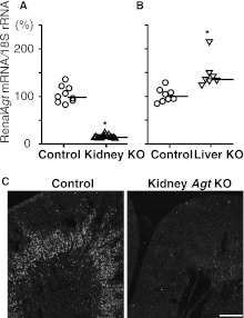
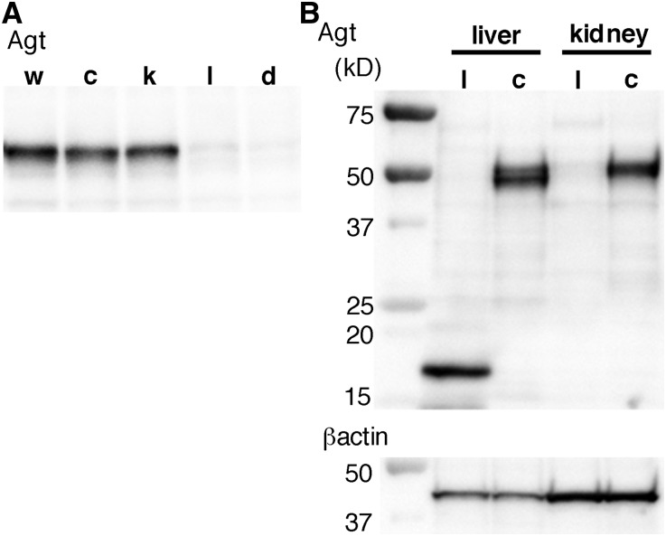
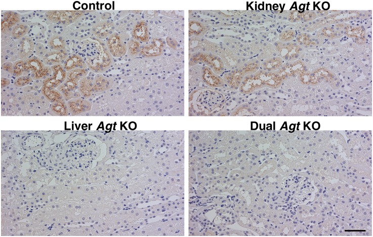
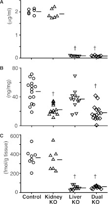
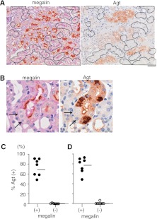
Angiotensin II content in the kidney is much higher than in the plasma, and it increases more in kidney diseases through an uncertain mechanism. Because the kidney abundantly expresses angiotensinogen mRNA, transcriptional dysregulation of angiotensinogen within the kidney is one potential cause of increased renal angiotensin II in the setting of disease. Here, we observed that kidney-specific angiotensinogen knockout mice had levels of renal angiotensinogen protein and angiotensin II that were similar to those levels of control mice. In contrast, liver-specific knockout of angiotensinogen nearly abolished plasma and renal angiotensinogen protein and renal tissue angiotensin II. Immunohistochemical analysis in mosaic proximal tubules of megalin knockout mice revealed that angiotensinogen protein was incorporated selectively in megalin-intact cells of the proximal tubule, indicating that the proximal tubule reabsorbs filtered angiotensinogen through megalin. Disruption of the filtration barrier in a transgenic mouse model of podocyte-selective injury increased renal angiotensin II content and markedly increased both tubular and urinary angiotensinogen protein without an increase in renal renin activity, supporting the dependency of renal angiotensin II generation on filtered angiotensinogen. Taken together, these data suggest that liver-derived angiotensinogen is the primary source of renal angiotensinogen protein and angiotensin II. Furthermore, an abnormal increase in the permeability of the glomerular capillary wall to angiotensinogen, which characterizes proteinuric kidney diseases, enhances the synthesis of renal angiotensin II.


Figures

Figure 1.
Figure 1.
Renal Agt mRNA in kidney Agt KO mice and liver Agt KO mice versus control mice. (A and B) Real-time RT-PCR analyses for Agt mRNA/18S rRNA. *P<0.05 compared with the control group. Bars represent median values. In kidney Agt KO mice, Agt mRNA is markedly low compared with control mice. In liver Agt KO mice, renal Agt mRNA is significantly increased. (C) In situ hybridization for Agt mRNA. In control kidneys, Agt mRNA is expressed in the outer stripe of the outer medulla. In kidney Agt KO mice, the signal is almost absent. Scale bar, 0.5 mm.
Figure 2.
Figure 2.
Western blot analyses for Agt protein. (A) In control (c) and kidney Agt KO mice (k), renal Agt protein content was not different from the content in wild-type mice (w). In contrast, both in liver (l) and dual (d) Agt KO mice, renal Agt protein is essentially absent; 25 μg protein were loaded in each lane. Control mice carry AgtloxP/loxP but not Cre gene. (B) In the liver of liver Agt KO mice, the ∼50-kD band is absent; instead, a smaller ∼16-kD band is present. Right two lanes again show near-complete absence of Agt protein in the kidney of liver Agt KO but not control mice; 10 μg hepatic protein or 15 μg renal protein was loaded.
Figure 3.
Figure 3.
Agt immunostaining in the kidney. In control mice, Agt protein is localized in proximal tubule cells. Unlike Agt mRNA, which is predominantly expressed in the S3 segment (Figure 1C), Agt protein is mainly localized in the S1 and S2 segments. The staining is in a granular pattern and distributes preferentially to the apical side of proximal tubule cells. Intensity of Agt staining is variable among nephrons (i.e., some nephrons contain more Agt protein than others). Within a given nephron, proximal tubule cells closer to Bowman’s space contain more Agt protein. These findings suggest that Agt protein in the proximal tubule cell represents reabsorption of Agt protein from the tubule fluid in a manner similar to the manner of albumin and many other proteins. In kidney Agt KO mice, Agt protein staining is similar to control mice in both intensity and pattern. In liver Agt KO mice, there is no Agt staining. Dual Agt KO mice are similar to liver Agt KO mice in this regard. Scale bar, 50 μm.
Figure 4.
Figure 4.
Plasma and urinary Agt protein and renal AII content in control, kidney Agt KO, liver Agt KO, and dual Agt KO mice. (A) Plasma Agt concentration is unaffected in kidney Agt KO mice and almost null in liver and dual Agt KO mice. (B) Urinary Agt to creatinine (Cr) ratio is significantly lower in kidney and dual Agt KO than control mice, but the ratio in liver Agt KO mice is not different from the ratio in control mice. (C) In kidney Agt KO mice, renal AII content is not different from the content in control. Contrastingly, it is markedly low in liver and dual Agt KO mice. Horizontal bars represent mean values in A and B and median in C. P<0.01 versus control mice.
Figure 5.
Figure 5.
Renal histology of Agt KO mice. Renal morphology is unaffected in control and kidney Agt KO mice. In liver and dual Agt KO mice, juxtaglomerular cells, afferent arterioles, and interlobular arteries are hypertrophic. Periodic acid-Schiff staining. Scale bar, 50 μm.
Figure 6.
Figure 6.
Agt protein reabsorption is dependent on megalin. (A) The left panel is megalin immunostaining in proximal tubule-specific mosaic megalin KO mouse kidney without podocyte injury. The proximal tubule cells outlined lack megalin staining. The adjacent section was stained for Agt (right panel). None of the outlined megalin KO cells are stained for Agt. Scale bar, 50 μm. (B) Serial kidney sections from NEP25/megalin KO mice 10 days after injection with LMB2 are stained for megalin and Agt. These pictures are taken from the outer stripe of outer medulla. Massive amounts of Agt protein are reabsorbed only in megalin-intact cells of the S3 segment but not megalin KO cells (arrows). Agt staining is in s granular pattern. Scale bar, 20 μm. (C and D) Agt positivity in mosaic megalin KO mice (D) with or (C) without glomerular sieving defect. The cortex that mainly contains S1 and S2 segments of the proximal tubule was photographed. Agt positivity was determined separately in megalin (+) and megalin proximal tubule cells (−). Regardless of the glomerular sieving conditions, megalin proximal tubule cells (−) did not contain Agt protein. Horizontal bars represent medians.
Figure 7.
Figure 7.
Sieving defect increases renal Agt protein and AII. NEP25 mice were injected with LMB2, an immunotoxin specific to hCD25, to induce sieving defect. Control NEP25 mice were injected with saline. (A) Western blot analysis for renal Agt protein. Kidneys of NEP25 mice with podocyte injury (+) contain more Agt protein than the kidneys of control mice (−). (B) Immunostaining for Agt protein. In kidneys with podocyte injury, Agt staining is enhanced and extended to the S3 segment. Scale bar, 100 μm. (C) Renal AII content. NEP25 mice with podocyte injury show significant increase in renal AII content. (D) Renal renin activity. NEP25 mice with podocyte injury show downregulated renin activity, reflecting sufficient volume repletion with plasma infusion in these nephrotic mice. Thus, the increase in renal AII found in these mice is not ascribed to increased renin. In C and D, data from individual mice are shown by open or closed circles. The median and 0.25 and 0.75 quantile values are shown by box plots placed next to the individual data. P<0.01 versus control NEP25 mice without podocyte injury.

Comment in

References

    1. Navar LG, Nishiyama A: Why are angiotensin concentrations so high in the kidney? Curr Opin Nephrol Hypertens 13: 107–115, 2004 - PubMed
    1. Kobori H, Nangaku M, Navar LG, Nishiyama A: The intrarenal renin-angiotensin system: From physiology to the pathobiology of hypertension and kidney disease. Pharmacol Rev 59: 251–287, 2007 - PubMed
    1. Pendergrass KD, Averill DB, Ferrario CM, Diz DI, Chappell MC: Differential expression of nuclear AT1 receptors and angiotensin II within the kidney of the male congenic mRen2. Lewis rat. Am J Physiol Renal Physiol 290: F1497–F1506, 2006 - PubMed
    1. Siragy HM, Carey RM: Role of the intrarenal renin-angiotensin-aldosterone system in chronic kidney disease. Am J Nephrol 31: 541–550, 2010 - PMC - PubMed
    1. Bader M, Ganten D: Update on tissue renin-angiotensin systems. J Mol Med (Berl) 86: 615–621, 2008 - PubMed

Publication types

MeSH terms