

Clinical outcome of patients with acute posterior circulation stroke and bilateral vertebral artery occlusion
- PMID: 22518265
- PMCID: PMC3317285
Clinical outcome of patients with acute posterior circulation stroke and bilateral vertebral artery occlusion
Abstract
Background and introduction: Patients presenting with posterior circulation acute ischemic events are occasionally noted to have occlusion of bilateral vertebral arteries with basilar artery blood flow entirely dependent from the anterior circulation. There is limited data about prognosis of such patients in literature.
Methods: Patients with acute posterior circulation ischemic stroke and bilateral vertebral artery occlusion (including contra-lateral hypoplastic vertebral artery without contribution to the basilar artery system) were identified prospectively from two academic centers. Data including clinical presentation, medical management, angiographic findings, recurrent events and outcome were collected and reported.
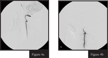
Results: A total of 4 patients presenting with acute ischemic events in the posterior circulation were identified to have bilateral vertebral artery occlusion at our center. One additional patient had a vertebral artery occlusion and a contra-lateral hypoplastic vertebral artery. In the functional evaluation of the blood flow with catheter angiography, the basilar artery was filling from the anterior circulation, with no antegrade flow from bilateral vertebral arteries injection in all 5 patients. Patients were treated with anti-platelets (n=4) or started on anti-coagulation after failing anti-platelet therapy (n=2). All patients had recurrent ischemic stroke with new ischemic lesions proven by diffusion weighted images on MRI within 2 to 70 days after the initial event.
Conclusion: Patients with acute posterior circulation ischemic stroke and bilateral vertebral artery occlusion are at high risk of having early recurrent ischemic events. Reestablishment of the antegrade vertebro-basilar blood flow through endovascular re-canalization might be an option to decrease stroke recurrence in selected patients with acute posterior circulation stroke and bilateral vertebral artery occlusion.
Keywords: bilateral; ischemic stroke; prognosis; vertebral artery occlusion.
Figures


Similar articles
-
A review of the diagnosis and management of vertebral basilar (posterior) circulation disease.Surg Neurol Int. 2018 May 24;9:106. doi: 10.4103/sni.sni_373_17. eCollection 2018. Surg Neurol Int. 2018. PMID: 29930872 Free PMC article. Review.
-
Occipital Artery to Extradural Vertebral Artery Bypass for Posterior Circulation Ischemia.Oper Neurosurg. 2019 May 1;16(5):527-538. doi: 10.1093/ons/opy143. Oper Neurosurg. 2019. PMID: 30982906
-
Childhood acute basilar artery thrombosis successfully treated with mechanical thrombectomy using stent retrievers: case report and review of the literature.Childs Nerv Syst. 2017 Feb;33(2):349-355. doi: 10.1007/s00381-016-3259-z. Epub 2016 Oct 4. Childs Nerv Syst. 2017. PMID: 27704247 Review.
-
Exploring vertebral artery stump syndrome: An overlooked cause of posterior ischemic strokes. A narrative review of current management options.J Stroke Cerebrovasc Dis. 2024 Aug;33(8):107819. doi: 10.1016/j.jstrokecerebrovasdis.2024.107819. Epub 2024 Jun 13. J Stroke Cerebrovasc Dis. 2024. PMID: 38878845 Review.
-
MRI in patients with acute basilar artery occlusion - DWI lesion scoring is an independent predictor of outcome.Int J Stroke. 2012 Jun;7(4):282-8. doi: 10.1111/j.1747-4949.2011.00705.x. Epub 2011 Dec 8. Int J Stroke. 2012. PMID: 22151607
Cited by
-
Patterns of ischemic posterior circulation strokes: A clinical, anatomical, and radiological review.Int J Stroke. 2022 Aug;17(7):714-722. doi: 10.1177/17474930211046758. Epub 2021 Sep 28. Int J Stroke. 2022. PMID: 34581223 Free PMC article. Review.
-
Rescue Endovascular Treatment to Prevent Neurological Deterioration in Acute Symptomatic Bilateral Vertebral Artery Occlusion.Neurointervention. 2023 Nov;18(3):182-189. doi: 10.5469/neuroint.2023.00381. Epub 2023 Oct 24. Neurointervention. 2023. PMID: 37871977 Free PMC article.
-
Acute Distal Vertebral Artery Occlusion in Patients with Asymmetrical Vertebral Artery Geometry: Role of Black-Blood-Enhanced MR Imaging.Diagnostics (Basel). 2022 Oct 1;12(10):2391. doi: 10.3390/diagnostics12102391. Diagnostics (Basel). 2022. PMID: 36292080 Free PMC article.
References
-
- Caplan LR. Bilateral distal vertebral artery occlusion. Neurology. 1983;33:552–558. - PubMed
-
- Bogousslavsky J, Gates PC, Fox AJ, Barnett HJ. Bilateral occlusion of vertebral artery: clinical patterns and long-term prognosis. Neurology. 1986;36:1309–1315. - PubMed
-
- Fisher CM. Occlusion of the vertebral arteries. Causing transient basilar symptoms. Arch Neurol. 1970;22:13–19. - PubMed
-
- Nakamura T, Yamamoto Y, Akiguchi I, Oiwa K, Nakajima K. [Bilateral vertebral artery occlusion] Rinsho Shinkeigaku. 1997;37:595–602. - PubMed
-
- Ruegg S, Engelter S, Jeanneret C, et al. Bilateral vertebral artery occlusion resulting from giant cell arteritis: report of 3 cases and review of the literature. Medicine (Baltimore) 2003;82:1–12. - PubMed
LinkOut - more resources
Full Text Sources