. 2012:2012:548453.
doi: 10.1155/2012/548453. Epub 2012 Feb 8.

Cataract surgery in uveitis

Affiliations

Cataract surgery in uveitis

Rupesh Agrawal et al. Int J Inflam. 2012.

Abstract

Cataract surgery in uveitic eyes is often challenging and can result in intraoperative and postoperative complications. Most uveitic patients enjoy good vision despite potentially sight-threatening complications, including cataract development. In those patients who develop cataracts, successful surgery stems from educated patient selection, careful surgical technique, and aggressive preoperative and postoperative control of inflammation. With improved understanding of the disease processes, pre- and perioperative control of inflammation, modern surgical techniques, availability of biocompatible intraocular lens material and design, surgical experience in performing complicated cataract surgeries, and efficient management of postoperative complications have led to much better outcome. Preoperative factors include proper patient selection and counseling and preoperative control of inflammation. Meticulous and careful cataract surgery in uveitic cataract is essential in optimizing the postoperative outcome. Management of postoperative complications, especially inflammation and glaucoma, earlier rather than later, has also contributed to improved outcomes. This manuscript is review of the existing literature and highlights the management pearls in tackling complicated cataract based on medline search of literature and experience of the authors.


Figures

Figure 1
Figure 1
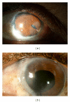
A 54-year-old farmer gave history of loss of vision and repeated episodes of redness and pain in the left eye following injury 40 years ago. B scan of the left eye showed findings suggestive of old vitreous hemorrhage and retinal detachment. Since visual acuity was doubtful perception of light, surgery was not advised for him. Diffuse (a) and slit (b) photographs show the left eye with peripheral corneal scar and peripheral anterior synechiae and total posterior synechia with cataract. At the temporal periphery, there is an incidental conjunctival lesion suggestive of actinic keratosis. Note the polychromatic crystals (cholesterolosis) deposited on the iris.
Figure 2
Figure 2
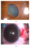
A 48-year-old lady was diagnosed as bilateral sclera-keratouveitis, with complicated cataract. She was investigated extensively and had a positive Mantoux test, for which she received anti-tubercular therapy. She underwent cataract surgery in the left eye with a preoperative visual acuity of counting fingers. Postoperatively, her visual acuity improved to 20/125, limited by the presence of a central corneal scar. (a) Shows the right eye showing evidence of healed scleritis, corectopia, and a complicated cataract. (b) Shows the left eye two months postoperatively.
Figure 3
Figure 3
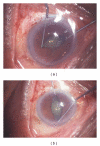
(a) Showing the eye with synechiae at papillary border, pigment deposition on the lens, and an early cataract and (b) showing presence of 360 degree posterior synechiae.
Figure 4
Figure 4
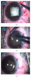
Intraoperative use of Kuglen hooks to stretch and dilate the pupils.
Figure 5
Figure 5
Postoperative slit lamp photograph showing minimally distorted pupil following pupil manipulation intraoperatively to negotiate posterior synechiae.
Figure 6
Figure 6
Intraoperative use of self-retaining iris hooks to dilate the pupils.
Figure 7
Figure 7
Postoperatively removal of self-retaining iris hooks.
Figure 8
Figure 8
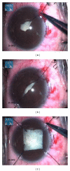
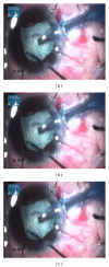
Intraoperative still video clip demonstrating the step of capsulorrhexis.
Figure 9
Figure 9
Intraoperative still surgical video clip showing the nucleus management in total white uveitic cataract.
Figure 10
Figure 10
Intraoperative still surgical video clip showing irrigation and aspiration of the soft lens matter.
Figure 11
Figure 11
A 45-year-old man, a treated case of Hansen's disease 15 years ago, presented with progressive visual loss. Examination showed active anterior uveitis and complicated cataract. He was treated with topical steroids and after 4 months, underwent phacoemulsification with PCIOL in the left eye. On the first postoperative day, his visual acuity improved to 20/50 (from preoperative vision of 20/200) with a mild fibrinous reaction in the anterior chamber. He returned to the emergency clinic on the next day with loss of vision and showed severe anterior chamber reaction with hypopyon. Vitreous appeared uninvolved. He was hospitalized and treated with intensive topical steroids and cycloplegics. He improved over the course of one week and regained good vision at the end of one month. (a, b) show the right and left eye with quiet anterior chambers and nondilating pupil with posterior synechia. (c, d) Diffuse and slit view of the left eye on the second postoperative day shows hypopyon and coagulum around the IOL. (e, f, g) Diffuse low and high magnification and slit view of the left eye two days after intensive treatment showing decrease in the inflammation. (h) The left eye shows near-quiet anterior chamber 2 weeks after treatment, the visual acuity has improved to 20/50.
Figure 12
Figure 12
A 36-year-old man presented with a history of redness and pain since two years and decreased vision since one year. Examination showed a total cataract in the right eye, with 360 degrees posterior synechiae. Investigations showed HLA-B 27 was positive. After the inflammation subsided, the patient underwent cataract surgery with synechiolysis. Postoperatively, there was increased anterior chamber inflammation, which was treated with oral and topical steroids. The patient regained visual acuity of 20/25; 3 months postoperatively and is on maintenance with oral methotrexate and topical steroids. (a, b) Diffuse and slit photograph of the right eye on the first postoperative day shows a membrane on the IOL which responded to intensive topical steroids and cycloplegics.

References

    1. Hooper PL, Rao NA, Smith RE. Cataract extraction in uveitis patients. Survey of Ophthalmology. 1990;35(2):120–144. - PubMed
    1. Malinowski SM, Pulido JS, Folk JC. Long-term visual outcome and complications associated with pars planitis. Ophthalmology. 1993;100(6):818–825. - PubMed
    1. Velilla S, Dios E, Herreras JM, Calonge M. Fuchs’ heterochromic iridocyclitis: a review of 26 cases. Ocular Immunology and Inflammation. 2001;9(3):169–175. - PubMed
    1. Foster CS, Rashid S. Management of coincident cataract and uveitis. Current Opinion in Ophthalmology. 2003;14(1):1–6. - PubMed
    1. Van Gelder RN, Leveque TK. Cataract surgery in the setting of uveitis. Current Opinion in Ophthalmology. 2009;20(1):42–45. - PubMed

LinkOut - more resources