Review
. 2012 May;18(5):273-82.
doi: 10.1016/j.molmed.2012.03.005. Epub 2012 Apr 19.

Protein misfolding and aggregation in cataract disease and prospects for prevention

Affiliations
Review

Protein misfolding and aggregation in cataract disease and prospects for prevention

Kate L Moreau et al. Trends Mol Med. 2012 May.

Abstract

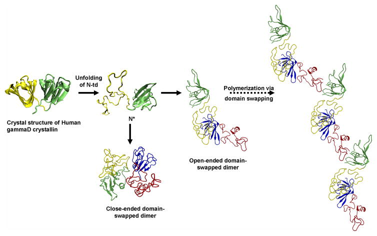
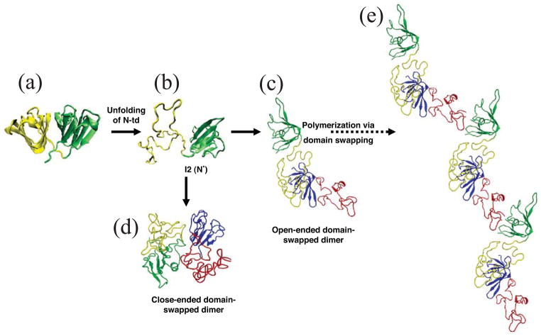
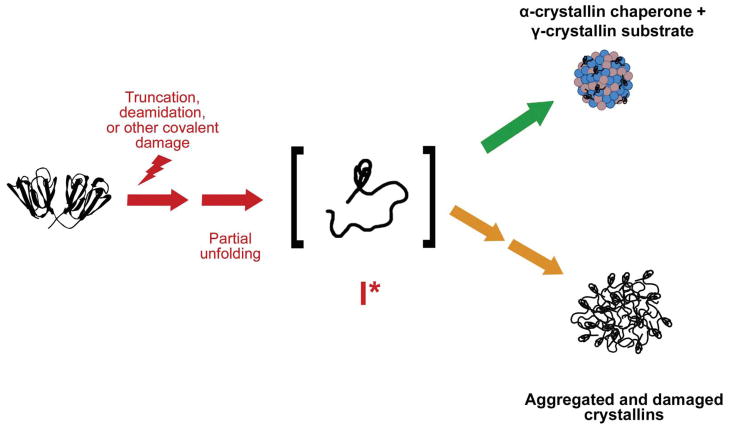
The transparency of the eye lens depends on maintaining the native tertiary structures and solubility of the lens crystallin proteins over a lifetime. Cataract, the leading cause of blindness worldwide, is caused by protein aggregation within the protected lens environment. With age, covalent protein damage accumulates through pathways thought to include UV radiation, oxidation, deamidation, and truncations. Experiments suggest that the resulting protein destabilization leads to partially unfolded, aggregation-prone intermediates and the formation of insoluble, light-scattering protein aggregates. These aggregates either include or overwhelm the protein chaperone content of the lens. Here, we review the causes of cataract and nonsurgical methods being investigated to inhibit or delay cataract development, including natural product-based therapies, modulators of oxidation, and protein aggregation inhibitors.


Figures

Figure 1
Figure 1
Structure of the lens and its major soluble proteins. (a) Cross section of the human lens highlighting the epithelial layer, immature and mature fiber cells. Reprinted with permission from [95]. Structures of the major soluble crystallins in human lenses: (b) cryo-EM structure of human αB-crystallin representative of the α-crystallin small heat shock protein (EMD-1776) [115]; (c) crystal structure of human βB2-crystallin representative of the β-crystallins (PDB ID 1YTQ) [116]; and (d) crystal structure of human γD-crystallin representative of the γ-crystallins (PDB ID 1HK0) [6]. Structures depicted in (b–d) were created using the program Chimera (http://www.cgl.ucsf.edu/chimera).
Figure 2
Figure 2
The prevalence of cataract in male and female populations. (a) Prevalence (per 100 people) in white individuals derived from four population-based studies. BDES: Beaver Dam Eye Study; BMES: Blue Mountains Eye Study; Melbourne VIP: Melbourne Visual Impairment Project; SEE Project: Salisbury Eye Evaluation Project. (b) Prevalence (per 100 people) in black individuals from two population-based studies. Reproduced with permission from [11].
Figure 3
Figure 3
Computational simulation of human γD-crystallin polymerization. (a) Crystal structure of human γD-crystallin. (b) Simulated monomeric aggregation precursor (I2), often referred as N* in the general mechanism of protein aggregation in literature. (c) Simulated structure of open-ended domain-swapped dimer. (d) Simulated structure of close-ended domain-swapped dimer. (e) Model of human γD-crystallin hexamer formed via domain swapping. Figure and legend reproduced with permission from [57].
Figure 4
Figure 4
Cataract viewed as a protein aggregation disease. Crystallin proteins, particularly in the lens nucleus, are present from birth and over time accumulate covalent modifications and damages resulting from proteolytic activity, non-enzymatic modifications, and oxidative damage. This leads to destabilization and partial unfolding of the polypeptide chains and a population of aggregation-prone intermediates. In young lenses, α-crystallin effectively recognizes and sequesters these destabilized intermediates (upper pathway). However, with age α-crystallin complexes become saturated with substrate and lens proteins are able to aggregate, resulting in light scatter and loss of visual acuity (lower pathway). Reprinted with permission from [54].

References

    1. Augusteyn RC. On the growth and internal structure of the human lens. Exp Eye Res. 2010;90:643–654. - PMC - PubMed
    1. Michael R, et al. Changes in the refractive index of lens fibre membranes during maturation--impact on lens transparency. Exp Eye Res. 2003;77:93–99. - PubMed
    1. Bassnett S. Lens organelle degradation. Exp Eye Res. 2002;74:1–6. - PubMed
    1. Delaye M, Tardieu A. Short-range order of crystallin proteins accounts for eye lens transparency. Nature. 1983;302:415–417. - PubMed
    1. Oyster CW. The human eye: structure and function. Sinauer Associates, Inc; 1999.

Publication types