Review
. 2012 Apr 20;36(4):515-28.
doi: 10.1016/j.immuni.2012.03.016.

Inborn errors of human JAKs and STATs

Affiliations
Review

Inborn errors of human JAKs and STATs

Jean-Laurent Casanova et al. Immunity. .

Abstract

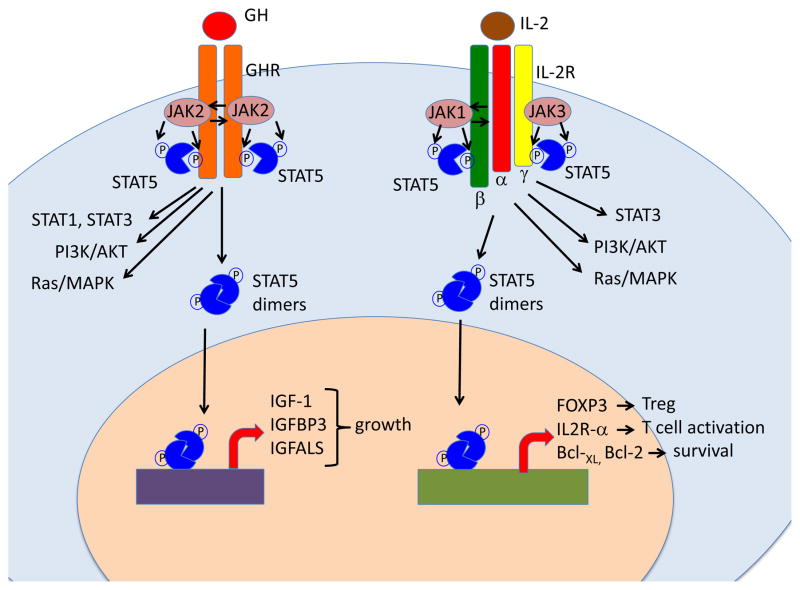
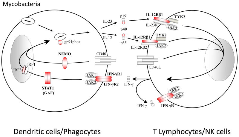
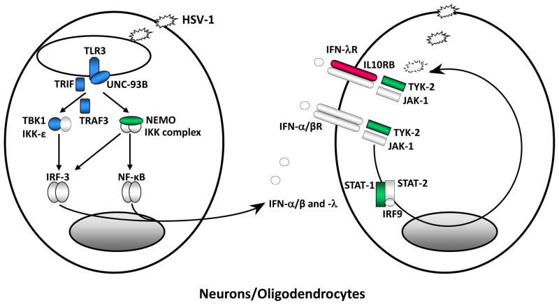
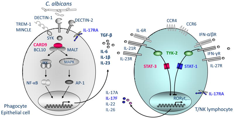
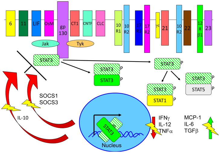
Inborn errors of the genes encoding two of the four human JAKs (JAK3 and TYK2) and three of the six human STATs (STAT1, STAT3, and STAT5B) have been described. We review the disorders arising from mutations in these five genes, highlighting the way in which the molecular and cellular pathogenesis of these conditions has been clarified by the discovery of inborn errors of cytokines, hormones, and their receptors, including those interacting with JAKs and STATs. The phenotypic similarities between mice and humans lacking individual JAK-STAT components suggest that the functions of JAKs and STATs are largely conserved in mammals. However, a wide array of phenotypic differences has emerged between mice and humans carrying biallelic null alleles of JAK3, TYK2, STAT1, or STAT5B. Moreover, the high degree of allelic heterogeneity at the human JAK3, TYK2, STAT1, and STAT3 loci has revealed highly diverse immunological and clinical phenotypes, which had not been anticipated.


Figures

Figure 1
Figure 1. Schematic representation of the role of JAK/STAT5 signaling in response to growth hormone and to interleukin-2
Left: Binding of growth hormone (GH) to GH receptor (GHR) homodimer triggers activation of the JAK2 kinase that phosphorylates the GHR, creating docking sites for STAT proteins. In the Figure, recruitment and JAK2-mediated phosphorylation of STAT5 is shown, but STAT1 and STAT3 are also activated. In addition, GH also triggers activation of the PI3K/AKT and Ras/MAPK pathways. Phosphorylated STAT5 proteins dimerize and translocate to the nucleus where they drive activation of target genes. Although both STAT5A and STAT5B are activated in response to GH, the genes that encode for Insulin Growth Factor-1 (IGF-1), IGF binding protein 3 (IGFBP3) and for the acid labile subunit of IGF-like binding protein (IGFALS) are under the direct control of STAT5B, and their expression is markedly repressed in STAT5B-mutated patients. This results in severe growth failure. Right: binding of interleukin-2 (IL-2) to its high-affinity receptor comprising IL-2Rα, -β and -γ chains promotes activation of JAK1 and JAK3 proteins, and recruitment of STAT3 and STAT5 to the phosphorylated IL-2R chains. PI3K/AKT and the Ras/MAPK pathways are also activated. STAT5A and STAT5B are phosphorylated and form homo- and hetero-dimers that translocate to the nucleus. Genes that are directly controlled by STAT5B and whose expression is significantly reduced in STAT5B-mutated patients include: FOXP3 (that promotes development and function of Treg cells), IL2RA (that favors T cell activation), and the genes that encodes for anti-apoptotic factors Bcl-2 and Bcl-XL. Failure to activate these genes in response to IL-2 explains the association of immunodeficiency and immune dysregulation in patients with STAT5B deficiency.
Figure 2
Figure 2. Inborn errors in the IL-12/23-IFN-γ pathway underlie Mendelian susceptibility to mycobacterial diseases (MSMD)
Schematic diagram of cytokine production and cooperation between phagocytes/dendritic myeloid cells and NK/T lymphocytes. The IL-12/IFN-γ circuit, the CD40/CD40L pathway and the oxidative burst (mediated in part by CYBB-encoded gp91, a component of the NADPH phagocyte oxydase) are crucial for protective immunity against mycobacterial infection in humans. Mutations in IFNGR1 or IFNGR2, encoding the ligand-binding and associated chains of the IFN-gR, impair cellular responses to IFN-g. Likewise, heterozygous dominant-negative mutations in STAT1 impair IFN-g but not IFN-a/b responses. Mutations in IL-12p40 or IL-12Rb1 impair IL-12-dependent induction of IFN-g. Mutations in CYBB that selectively impair the respiratory burst in monocyte-derived macrophages are associated with MSMD. Heterozygous dominant-negative mutations in IRF8 impair the development of IL-12-producing CD1cCD11c DCs. Proteins for which mutations in the corresponding genes have been identified and associated with MSMD, are shown in red. The allelic heterogeneity is described in Table 1.
Figure 3
Figure 3. Inborn errors of TLR3-dependent, IFN-a/b and -l immunity underlie childhood herpes simplex virus 1 encephalitis (HSE)
Schematic representation of the production of and response to IFN-α/-β, and IFN-λ in anti-HSV-1 immunity in the central nervous system (CNS), based on the genetic dissection of children with HSE. Like most viruses HSV-1 produce dsRNA intermediates during its replication. TLR3 is an endosomal transmembrane receptor for dsRNA. The recognition of dsRNA by TLR3 induces activation of the IRF-3 and NF-kB pathways via TRIF, leading to IFN-α/-β and/or IFN-λ production. TLR3, UNC-93B, TRIF, TRAF3, TBK1 and NEMO deficiencies are all associated with impaired IFN-α/-β and/or IFN-λ production and predisposition to HSE in the course of primary infection by HSV-1. The binding of IFN-α/β and IFN-λ to their receptors induce the phosphorylation of JAK1 and TYK-2, activating the signal transduction proteins STAT-1, STAT-2 and IRF9. This complex is translocated as a heterotrimer to the nucleus, where it acts as a transcriptional activator, binding to specific DNA response elements in the promoter region of IFN-inducible genes. STAT-1 and TYK2 deficiencies are associated with impaired IFN-α/β responses and, for STAT1, impaired IFN-λ responses and predisposition to HSE. Proteins for which genetic mutations have been identified and associated with susceptibility to isolated HSE are shown in blue. Proteins for which genetic mutations have been identified and associated with susceptibility to mycobacterial, bacterial and viral diseases, including HSE, are shown in green. Proteins for which genetic mutations have been identified but not associated with susceptibility to infectious diseases are shown in red. This figure will be revised as new results are obtained with the genetic and immunological dissection of children with HSE and other viral diseases.
Figure 4
Figure 4. Inborn errors of IL-17 immunity underlie chronic mucocutaenous candidiasis (CMC)
Upon C. albicans recognition via various cell surface receptors, adaptor molecules SYK and CARD9 mediate the induction of pro-inflammatory cytokines by myeloid and epithelial cells. Pro-inflammatory cytokines, such as IL-6, IL-21 or IL-23, activate T lymphocytes via STAT3 resulting in their differentiation into IL-17-producing T cells. These cells constitute a major component of the immune defense against C. albicans, as mutations in IL-17F or IL-17RA underlie CMC. Gain of function mutations in STAT1 inhibit this differentiation by mechanisms that have remained elusive. Enhanced stimulation via IFN-a/b, IFN-g, IFN-d and IL-27 might be responsible for this phenotype. The molecule TYK2 is known to act upstream of STAT1 and STAT3. It is unclear whether patients with AR TYK2 deficiency display CMC. TYK2 deficient patients. Proteins represented in red are mutated in patients with CMC only. Proteins represented in blue are mutated in patients with CMC and other infections.
Figure 5
Figure 5. Multiple receptors activate the STAT3 signaling pathway
The signaling and inhibitions of STAT3 are shown, with areas of special emphasis for the dominant negative mutants shown. Crosshatched STAT3 depicts the AD-mutant form of the molecule, with each heterodimer in which it participates being inhibited from function. The thunderbolts are to indicate where a normal function is being inhibited. STAT3 is both involved in IL-10 signal transduction and in IL-10 expression, both of which are affected in Job’s syndrome.

References

    1. O’Shea JJ, Gadina M, Kanno Y. Cytokine signaling: birth of a pathway. J Immunol. 2011;187:5475–5478. doi: 10.4049/jimmunol.1102913. 187/11/5475 [pii] - DOI - PMC - PubMed
    1. Farrar JD, et al. Selective loss of type I interferon-induced STAT4 activation caused by a minisatellite insertion in mouse Stat2. Nat Immunol. 2000;1:65–69. - PubMed
    1. Casanova JL, Abel L. The human model: a genetic dissection of immunity to infection in natural conditions. Nat Rev Immunol. 2004;4:55–66. - PubMed
    1. Glanzmann E, Riniker P. [Essential lymphocytophthisis; new clinical aspect of infant pathology] Annales paediatrici International review of pediatrics. 1950;175:1–32. - PubMed
    1. Gennery AR, et al. Transplantation of hematopoietic stem cells and long-term survival for primary immunodeficiencies in Europe: entering a new century, do we do better? J Allergy Clin Immunol. 2010;126:602–610. e601–611. doi: 10.1016/j.jaci.2010.06.015. S0091-6749(10)00992-9 [pii] - DOI - PubMed

Publication types

MeSH terms