

Review
doi: 10.1016/j.hoc.2012.02.002.
The antifolates
Affiliations
- PMID: 22520983
- PMCID: PMC3777421
- DOI: 10.1016/j.hoc.2012.02.002
Item in Clipboard
Review
The antifolates
Hematol Oncol Clin North Am.
2012 Jun.
Abstract
This article focuses on the cellular, biochemical, and molecular pharmacology of antifolates and how a basic understanding of the mechanism of action of methotrexate, its cytotoxic determinants, mechanisms of resistance, and transport into and out of cells has led to the development of a new generation of antifolates, a process that continues in the laboratory and in the clinics. New approaches to folate-based cancer chemotherapy are described based on the targeted delivery of drugs to malignant cells.
Copyright © 2012 Elsevier Inc. All rights reserved.
Figures
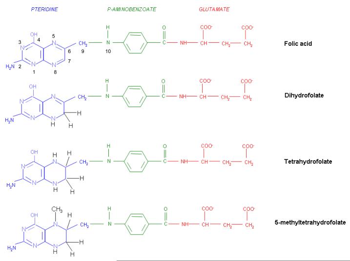
The structures of folic acid, dihydrofolate, tetrahydrofolate, and 5-methyltetrahydrofolate. Folic acid is not a physiological folate but is an important source of folate in foods and vitamins. Within cells folic acid is reduced to dihydrofolate by dihydrofolate reductase (DHFR-see Figure 2), albeit at a very slow rate since it is a very poor substrate for this enzyme. Dihydrofolate is the major oxidized form of folates within cells, and the preferred substrate for DHFR, mediating the formation of tetrahydrofolate that goes on to form a variety of tetrahydrofolate cofactors. Seen here is the major dietary folate, and the major folate in the blood, 5-methyltetrahydrofolate.
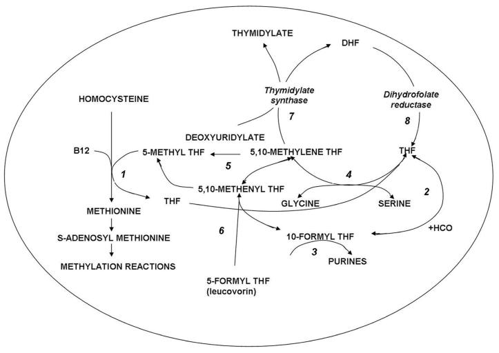
Folate-dependent reactions within cells. Reaction1: 5-methyltetrahydrofolate (5-methylTHF) enters the folate cycle with the provision of its methyl group to homocysteine in the synthesis of methionine, a vitamin B12 dependent reaction mediated by methionine synthase. The tetrahydrofolate (THF) moiety can then acquire a carbon at various oxidation states. Reaction 2: Formate is added at the N10 position to form 10-formylTHF which provides two carbons in Reaction 3 for the synthesis of purines. Reaction 4: 5,10-methyleneTHF is formed from serine and THF in a reaction mediated by serine hydroxyl methyltransferase. Reaction 5: 5-10-methyleneTHF is reduced irreversibly to 5-methylTHF. Reaction 6: 5-formytetrahydrofolate dehydrase is the mechanism by which 5-formylTHF (leucovorin) enters these cyclical folate pathways. Reaction 7: The formation of thymidylate mediated by thymidylate synthase; the THF moiety is oxidized to dihydrofolate (DHF). Reaction 8: DHF is reduced to THF by dihydrofolate reductase. The latter is essential for maintaining THF cofactor pools within mammalian cells.
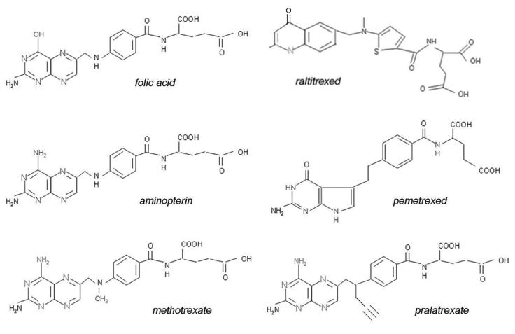
The structures of folic acid and a group of antifolates; except for aminopterin, all are in clinical use in the United States and/or elsewhere.
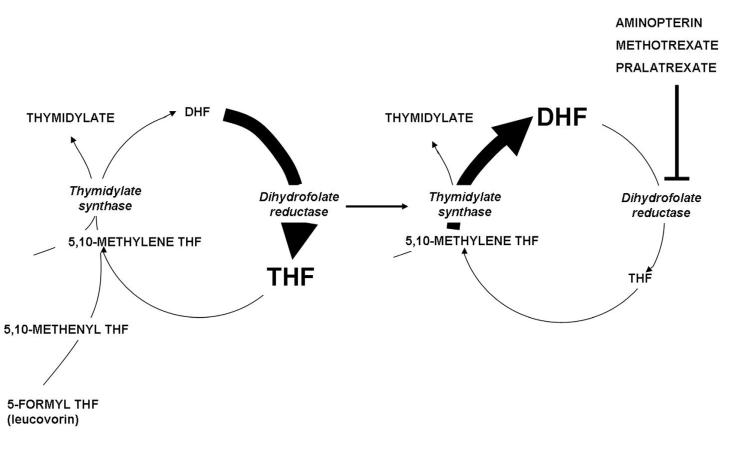
The impact of dihydrofolate reductase inhibitors on folate pools within cells. The left panel illustrates that under physiological conditions dihydrofolate (DHF) produced during the synthesis of thymidylate mediated by thymidylate synthase is reduced to tetrahydrofolate (THF) so rapidly due to the high levels of dihydrofolate reductase (DHFR) within cells that the DHF level is trivial compared to the level of tetrahydrofolate. The right panel illustrates the impact of suppression of DHFR by several 4-amino antifolates. High levels of DHF build up in cells by continued thymidylate synthase activity, interconversion of THF cofactors to 5,10-methyleneTHF and oxidation to DHF. This leads to depletion of THF cofactor levels within cells and cessation of THF cofactor-dependent reactions. High DHF levels compete with MTX for the small percentage of enzyme sufficient to maintain THF cofactor pools within cells.
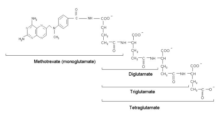
The structure of the polyglutamate derivatives of MTX. Glutamates are progressively added to the γ-carboxyl of the MTX molecule and each successive polyglutamate derivative.
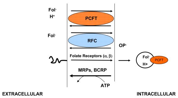
Folate-specific transport routes in mammalian cells. There are two carrier mechanisms: the reduced folate carrier (RFC), an organic phosphate (OP−) antiporter, and the proton-coupled folate transporter (PCFT), a proton-folate symporter. Both carriers transport folates and antifolates into cells against an electrochemical-potential gradient. Multidrug resistance-associated proteins (MRPs) and the breast cancer resistance protein (BCRP) utilize the energy released in the hydrolysis of ATP to pump folate and antifolate monoglutamates out of cells. BCRP and some MRPs can export lower polyglutamates as well. Folate receptors transport folates into cells by an endocytic mechanism. Folate export from acidified endosomes is mediated, in part, by PCFT.
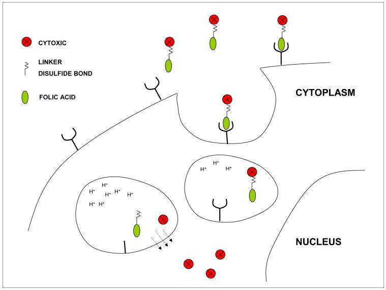
Receptor mediated endocytosis of folic acid linked to cytotoxic agents. A drug is coupled to folic acid via a hydrophilic linker molecule and a segment containing a cleavable disulfide bond. The complex binds to the folate receptor at the cell membrane which invaginates and forms a vesicle that circulates within the endosomal compartment. As the vesicle matures the reducing potential increases, rupturing the disulfide bond releasing the cytotoxic which diffuses out of the endosome to reach its intracellular target. Folic acid can also be linked to Technicium-99 to establish the presence of folate receptors and the competence of the endocytic mechanism before treatment with the cytotoxic conjugate.
References
-
- Skipper HE, Perry S. Kinetics of normal and leukemic leukocyte populations and relevance to chemotherapy. Cancer Res. 1970;30:1883–97. - PubMed
-
- Goldin A, Venditti JM, Humphreys SR, et al. A quantitative comparison of the antileukemic effectiveness of two folic acid antagonists in mice. J Natl Cancer Inst. 1955;15:1657–64. - PubMed
-
- Farber S, Diamond LK, Mercer RD, et al. Temporary remission in acute leukemia in children produced by folic acid antagonist, 4-aminopteroyl glutamic acid (aminopterin) N Engl J Med. 1948;238:787–93. - PubMed
-
- Goldman ID, Chattopadhyay S, Zhao R, et al. The Antifolates: Evolution, New Agents in the Clinic, and How targeting delivery via specific membrane transporters is driving the development of a next generation of folate analogs. Curr Opin Investig Drugs. 2010;11:1409–23. - PubMed
-
- Chattopadhyay S, Moran RG, Goldman ID. Pemetrexed: biochemical and cellular pharmacology, mechanisms, and clinical applications. Mol Cancer Ther. 2007;6:404–17. - PubMed
Publication types
MeSH terms
Substances
Grants and funding
LinkOut - more resources
Full Text Sources
Other Literature Sources
