. 2012 Mar 4;4(5):375-82.
doi: 10.1038/nchem.1290.

Metal-directed, chemically tunable assembly of one-, two- and three-dimensional crystalline protein arrays

Affiliations

Metal-directed, chemically tunable assembly of one-, two- and three-dimensional crystalline protein arrays

Jeffrey D Brodin et al. Nat Chem. .

Abstract

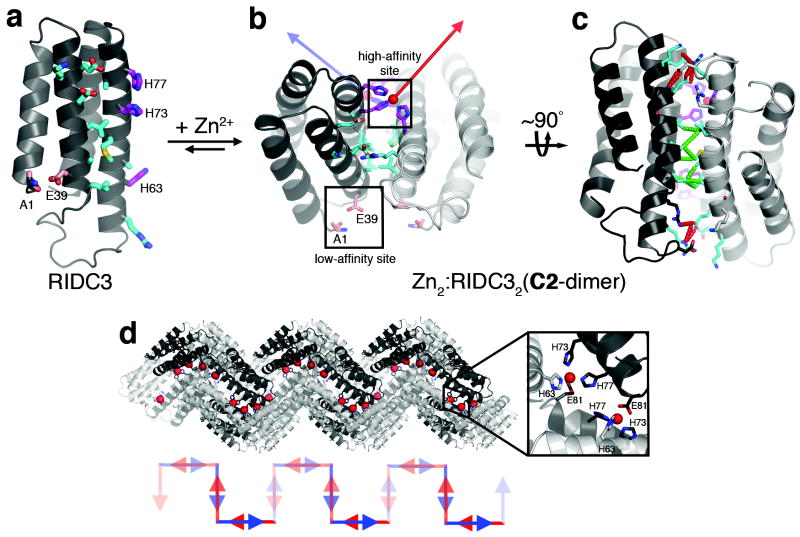
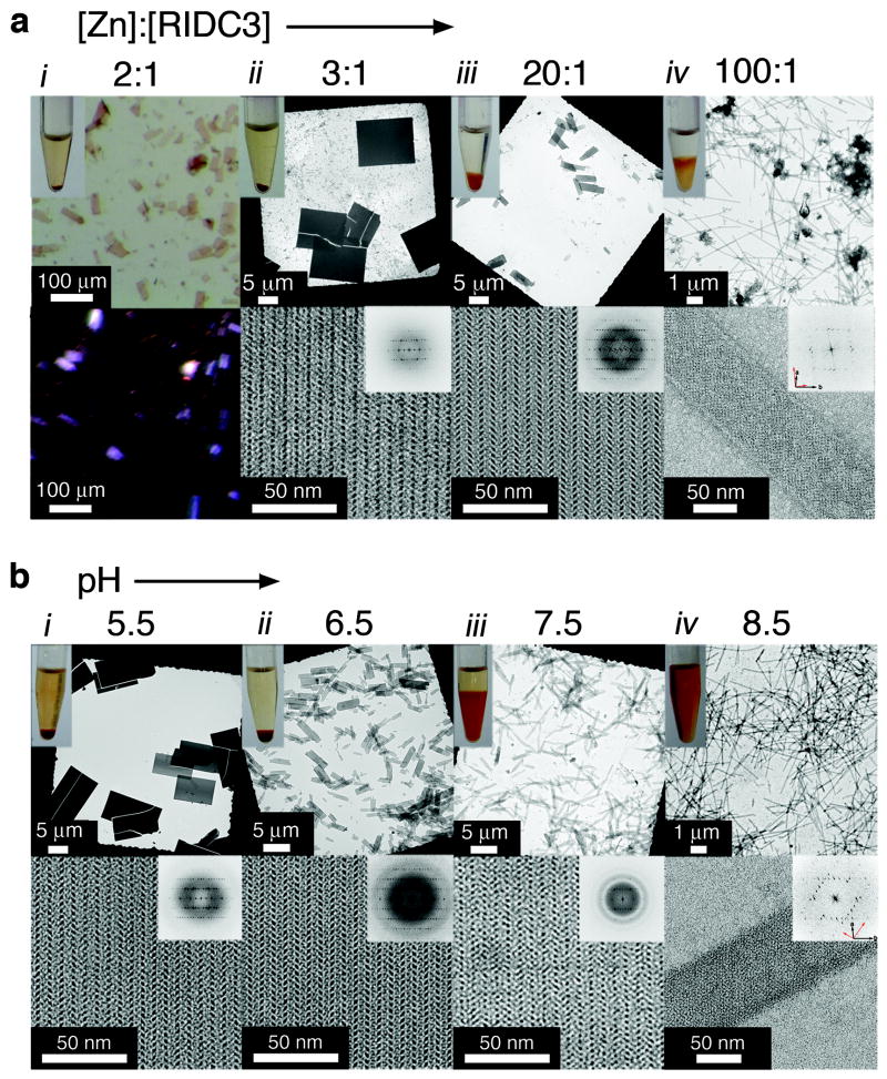
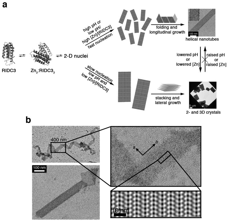
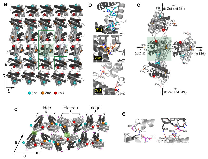
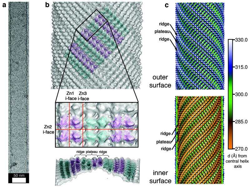
Proteins represent the most sophisticated building blocks available to an organism and to the laboratory chemist. Yet, in contrast to nearly all other types of molecular building blocks, the designed self-assembly of proteins has largely been inaccessible because of the chemical and structural heterogeneity of protein surfaces. To circumvent the challenge of programming extensive non-covalent interactions to control protein self-assembly, we have previously exploited the directionality and strength of metal coordination interactions to guide the formation of closed, homoligomeric protein assemblies. Here, we extend this strategy to the generation of periodic protein arrays. We show that a monomeric protein with properly oriented coordination motifs on its surface can arrange, on metal binding, into one-dimensional nanotubes and two- or three-dimensional crystalline arrays with dimensions that collectively span nearly the entire nano- and micrometre scale. The assembly of these arrays is tuned predictably by external stimuli, such as metal concentration and pH.


Conflict of interest statement

The authors declare no competing financial interests.

Figures

Figure 1
Figure 1. Design and initial crystallographic characterization of RIDC3
a, Cartoon representation of the RIDC3 monomer. Surface residues predicted by RosettaDesign to stabilize the Zn-induced RIDC3 dimer, the high-affinity and the low-affinity Zn binding sites are highlighted as cyan, magenta and pink sticks, respectively. b, Zn binding by RIDC3 produces a C2-symmetric dimer (Zn2:RIDC32 or C2-dimer) with two orthogonal coordination vectors (red and blue arrows) originating from the two high-affinity Zn sites. The C2-dimer geometry is derived from the structure of the Zn-mediated tetramer of MBPC1 (Zn4:MBPC14) as illustrated in Supplementary Fig. 1. c, Rotated view of the C2-dimer showing the crystallographically observed hydrophobic (dashed green lines) and polar (dashed red lines) interactions in the dimeric interface. d, Pairwise Zn coordination interactions between neighboring C2-dimers involving the high-affinity sites and E81 (inset) in the lattice leads to a helical 1D chain. Shown below is a cartoon representation of the C2-dimer units in this chain, illustrated as pairs of blue and red arrows that approximately represent the direction of Zn coordination vectors.
Figure 2
Figure 2. Zn-induced RIDC3 self-assembly in solution characterized by light and electron microscopy
a, Dependence of the morphology of self-assembled RIDC3 arrays on the [Zn]:[RIDC3] ratio at [RIDC3]=50 μM and pH=5.5. b, Dependence of the morphology of self-assembled RIDC3 arrays on pH at [RIDC3]=100 μM and [Zn]=300 μM. These microscopy images illustrate a trend of decreasing array size with either an increase in the [Zn]:[RIDC3] ratio (a) or an increase in pH (b). For [Zn]:[RIDC3]=2 at pH=5.5 (column i in a), only macroscopic crystalline arrays were observed and were viewed by light microscopy with (top) and without (bottom) polarizers. The birefringence of the RIDC3 arrays is indicative of crystalline order. In all other experiments, the top and the bottom rows show low and high magnification TEM images, respectively, with the computed Fourier transforms of the high-magnification images shown as insets. Discrete reflections in the Fourier transforms with identical spacings under all conditions demonstrate that all RIDC3 arrays are crystalline and contain the same underlying two-dimensional lattice. The overlapping lattice axes in the tubular structures (column iv) are highlighted with black and red arrows (bottom right of transform).
Figure 3
Figure 3. Model for RIDC3 self-assembly
a, Hypothetical model for Zn-mediated RIDC3 self-assembly under fast and slow nucleation conditions. Fast nucleation/growth conditions (high pH or high [Zn]:[RIDC3] ratio) promote the formation of many 2D nuclei, which “roll up” into helical nanotubes. Under slow nucleation conditions (low pH and low [Zn]:[RIDC3]), a smaller number of large 2D nuclei form, which can stack up in the third dimension due to the lack of repulsive interactions near the isoelectric point (pI = 5.3) of RIDC3. b, TEM images of a 2D RIDC3 ribbon and a tubular structure with frayed ends, illustrating the interconversion between tubular and sheet-like morphologies. Shown in the right bottom corner is the 2D image reconstruction of a single-layered portion of the ribbon, indicating the molecular arrangement of RIDC3 molecules.
Figure 4
Figure 4. Structural basis of Zn-mediated RIDC3 assembly
a, Crystallographically-determined molecular arrangement in 2D Zn-RIDC3 sheets, viewed normal to the bc plane of 3D crystalline arrays. A single C2-dimer is highlighted in the shaded green box. b, Close-up view of the three different Zn coordination environments that enable RIDC3 self-assembly in two dimensions. Zn1 and Zn2 sites are formed by the high affinity coordination motif described in Fig. 1, whereas Zn3 is formed by the low affinity coordination motif. c, Close-up view of the T-shaped boxed area in (a). d, Contacts between each 2D RIDC3 layer, responsible for growth in the third dimension, are highlighted in shaded green boxes and detailed in e.
Figure 5
Figure 5. RIDC3 nanotube structure and assembly
a, A typical vitrified, unstained nanotube used for helical cryo-EM reconstruction; see Supplementary Fig. S14 for collective views of RIDC3 nanotubes. b, Tetrameric RIDC3 units (Zn1-linked dimer of C2-dimers) modeled into the reconstructed tube density map. (top) View of the outer surface showing the alternating orientations (cyan and magenta) of tetrameric units; (middle) closeup view of tube surface with modeled tetramers; (bottom) lateral view of the tube highlighting its curvature. c, Radially color-coded representations of the outer (top) and inner (bottom) surfaces of the reconstructed nanotube revealing ridges and plateaus consistent with the X-ray crystal structure.
Figure 6
Figure 6. Rhodamine-directed stacking of 2D RIDC3 arrays
a, Structural model of a 2D RIDC3 sheet uniformly labeled with Rhodamine Red C2 (green sticks) at Cys21. b and c, TEM images of fully matured, negatively stained rhodamine-C21RIDC3 crystals obtained at pH 8.5 (b) and pH 5.5 (c). The formation of 3D structures at pH 8.5 rather than nanotubes evidences that rhodamine labels promote favorable interactions between 2D, Zn-mediated RIDC3 layers. d, UV-visible absorption spectrum of a 50 μM rhodamine-C21RIDC3 (1-mm pathlength) sample obtained prior to Zn addition (black), after crystal maturation (red) and upon dissolution of crystals by addition of EDTA (green). The intense band at 415 nm is due to the haem Soret absorption. e, CD spectra of the same rhodamine-C21RIDC3 sample. The increase in absorption at 534 nm upon Zn addition (d) indicates the formation of rhodamine dimers, while the emergence of a CD signal in the 520-630 nm region (e) during the maturation of rhodamine-C21RIDC3 crystals suggests that the rhodamine dimers are in discrete, rigid environments.

Comment in

References

    1. Mann S. Life as a nanoscale phenomenon. Angew Chem Int Ed Eng. 2008;47:5306–5320. - PubMed
    1. Nguyen ST, Gin DL, Hupp JT, Zhang X. Supramolecular chemistry: Functional structures on the mesoscale. Proc Natl Acad Sci USA. 2001;98:11849–11850. - PMC - PubMed
    1. Seeman NC, Belcher AM. Emulating biology: Building nanostructures from the bottom up. Proc Natl Acad Sci USA. 2002;99:6451–6455. - PMC - PubMed
    1. Shenton W, Pum D, Sleytr UB, Mann S. Synthesis of cadmium sulphide superlattices using self-assembled bacterial S-layers. Nature. 1997;389:585–587.
    1. McMillan RA, Paavola CD, Howard J, Chan SL, Zaluzec NJ, Trent JD. Ordered nanoparticle arrays formed on engineered chaperonin protein templates. Nat Mater. 2002;1:247–252. - PubMed

Publication types