. 2012 May;44(5):581-5.
doi: 10.1038/ng.2253.

Mutations in ISPD cause Walker-Warburg syndrome and defective glycosylation of α-dystroglycan

Affiliations

Mutations in ISPD cause Walker-Warburg syndrome and defective glycosylation of α-dystroglycan

Tony Roscioli et al. Nat Genet. 2012 May.

Abstract

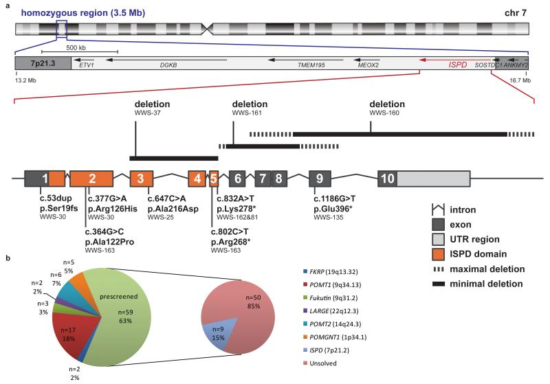
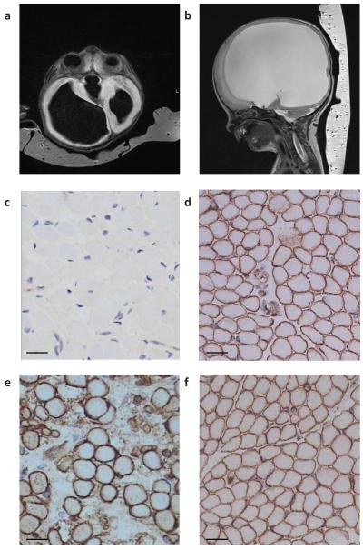
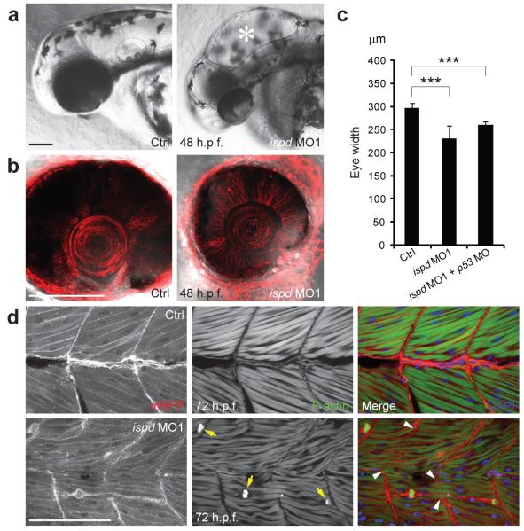
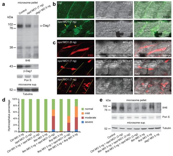
Walker-Warburg syndrome (WWS) is an autosomal recessive multisystem disorder characterized by complex eye and brain abnormalities with congenital muscular dystrophy (CMD) and aberrant a-dystroglycan glycosylation. Here we report mutations in the ISPD gene (encoding isoprenoid synthase domain containing) as the second most common cause of WWS. Bacterial IspD is a nucleotidyl transferase belonging to a large glycosyltransferase family, but the role of the orthologous protein in chordates is obscure to date, as this phylum does not have the corresponding non-mevalonate isoprenoid biosynthesis pathway. Knockdown of ispd in zebrafish recapitulates the human WWS phenotype with hydrocephalus, reduced eye size, muscle degeneration and hypoglycosylated a-dystroglycan. These results implicate ISPD in a-dystroglycan glycosylation in maintaining sarcolemma integrity in vertebrates.


Figures

Figure 1
Figure 1
Overview of genetic data in the patient cohort. (a) Schematic representation of the intragenic deletions, point mutations and homozygosity mapping data from WWS families with ISPD mutations. Ideogram of chromosome 7 showing the 3.5 Mb region of common homozygosity at band 7p21.3 flanked by SNPs rs194034 and rs818323 that was identified in family WWS-25. The position of three partially overlapping intragenic deletions in ISPD is indicated above the intron-exon structure of the gene. At the bottom the position of homozygous and compound heterozygous mutations is shown with respect to the ISPD protein domain structure. (b) Identified mutations in our total WWS/MEB cohort in number and percentage per gene. 94 families were available for research and prescreening revealed mutations in one of the six known genes in 35 families.
Figure 2
Figure 2
MR images and muscle staining of patient WWS-160. (a) Axial T1 weighted and (b) parasagittal T2 cerebral MRIs showing hydrocephalus. (c) Muscle biopsy showed almost absent αDG glycosylation using IIH6 antibody in muscle in comparison to (d) IIH6 staining in a normal control muscle biopsy. (e) Spectrin staining in the patient was not visibly different from (f) normal control spectrin staining. Scale bars, 20 μm.
Figure 3
Figure 3
Knockdown of zebrafish ispd recapitulates pathological defects of human WWS. (a) Compared with uninjected controls, zebrafish embryos injected with ispd MO1 (7 ng) showed characteristic hydrocephalus (asterisk) by 48 h.p.f. Scale bar, 100 μm. (b) Embryos injected with ispd MO1 (7 ng) showed microphthalmia by 48 h.p.f. in comparison to controls; cell membranes were visualized by membrane-localized red fluorescent protein (mRFP). Scale bar, 100 μm. (c) Eye width measurements in control (297.52±9.06 μm, n=25) and ispd MO1 (7 ng) injected embryos (230.8±28.35 μm, n=25; ***P= 4.68E-12). Co-injection of p53 MO (6 ng) with ispd MO1 (6 ng) still resulted in reduced eye size (260.28 ± 6.86 μm, n=25; ***P= 1.39E-20), suggesting that this phenotype was not a consequence of MO off-target effects mediated by p53-induced cell death. Error bars indicate s.d. (d) Control embryos display intact muscle fibers that anchor to chevron-shaped MTJ. Embryos injected with ispd MO1 (7 ng) showed muscle fiber degeneration by 72 h.p.f. Retracting muscle fibers were revealed by condensed F-actin (arrows) and collapsed sarcolemma (visualized by mRFP). Abnormally elongated muscle fibers spanned disrupted MTJ (arrowheads) in zebrafish embryos lacking Ispd. DAPI indicates nuclei. Scale bar, 100 μm.
Figure 4
Figure 4
Hypoglycosylation of αDG and disrupted sarcolemma integrity in ispd MO1-injected zebrafish embryos. (a) Western blot analysis of microsome pellets and supernatant from control, ispd MO1 (7 ng) and dag1 MO (5 ng) injected embryos at 48 h.p.f. Compared with control embryos, ispd MO1-injected embryos showed a reduction of glycosylated αDG (IIH6; 76-102 kDa) with a slight decrease of ßDG, which is probably a secondary reduction due to protein instability caused by defective glycosylation of αDG as reported previously,. Both glycosylated αDG and ßDG were almost absent in dag1 MO-injected embryos. Equal protein loading was demonstrated by Ponceau S (PonS) staining and unknown glycoproteins detected by IIH6 antibody in all three lanes (<38 kDa). Equivalent amounts of γ- and acetylated tubulins were detected in corresponding microsome supernatant. (b) Laminins remained localized at the MTJ in ispd MO1-injected embryos (7 ng). Positive fluorescent signal within degenerated muscle fibers (arrows) was probably due to disrupted sarcolemma integrity. Scale bar, 50 μm. (c) MTJ-anchored muscle fibers were infiltrated by EBD in ispd MO1-injected embryos before the onset of muscle degeneration. Dashed lines indicate MTJ. DIC, differential interference contrast microscopy; ANT, anterior myotome; PST, posterior myotome. Scale bar, 50 μm. (d) Injection of sub-effective doses of ispd, fktn, fkrp and control MO together or alone. Increase in the percentage of embryos with hydrocephalus suggests genetic interactions between ispd, fktn and fkrp. Each bar represents a combination of two independent experiments, scored blindly according to criteria exemplified in Supplementary Fig. 8a. n=94–139 embryos. (e) Western blotting with IIH6 antibody showed a reduction of glycosylated αDG in embryos co-injected with ispd MO1 and fktn/fkrp MO as compared to control MO and ispd MO1 co-injected embryos, and single fktn or fkrp MO-injected embryos. As a negative control, almost absent αDG glycosylation is shown for dag1 MO injected embryos.

References

    1. van Reeuwijk J, Brunner HG, van Bokhoven H. Glyc-O-genetics of Walker-Warburg syndrome. Clin Genet. 2005;67:281–9. - PubMed
    1. Kobayashi K, et al. An ancient retrotransposal insertion causes Fukuyama-type congenital muscular dystrophy. Nature. 1998;394:388–92. - PubMed
    1. Yoshida A, et al. Muscular dystrophy and neuronal migration disorder caused by mutations in a glycosyltransferase, POMGnT1. Dev Cell. 2001;1:717–24. - PubMed
    1. Beltran-Valero de Bernabe D, et al. Mutations in the O-mannosyltransferase gene POMT1 give rise to the severe neuronal migration disorder Walker-Warburg syndrome. Am J Hum Genet. 2002;71:1033–43. - PMC - PubMed
    1. Brockington M, et al. Mutations in the fukutin-related protein gene (FKRP) cause a form of congenital muscular dystrophy with secondary laminin alpha2 deficiency and abnormal glycosylation of alpha-dystroglycan. Am J Hum Genet. 2001;69:1198–209. - PMC - PubMed

Publication types