. 2012 Jul;61(7):1752-9.
doi: 10.2337/db11-1520. Epub 2012 Apr 20.

Circulating preproinsulin signal peptide-specific CD8 T cells restricted by the susceptibility molecule HLA-A24 are expanded at onset of type 1 diabetes and kill β-cells

Affiliations

Circulating preproinsulin signal peptide-specific CD8 T cells restricted by the susceptibility molecule HLA-A24 are expanded at onset of type 1 diabetes and kill β-cells

Deborah Kronenberg et al. Diabetes. 2012 Jul.

Abstract

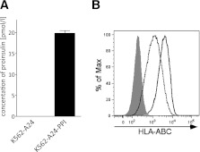
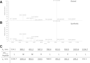
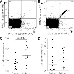
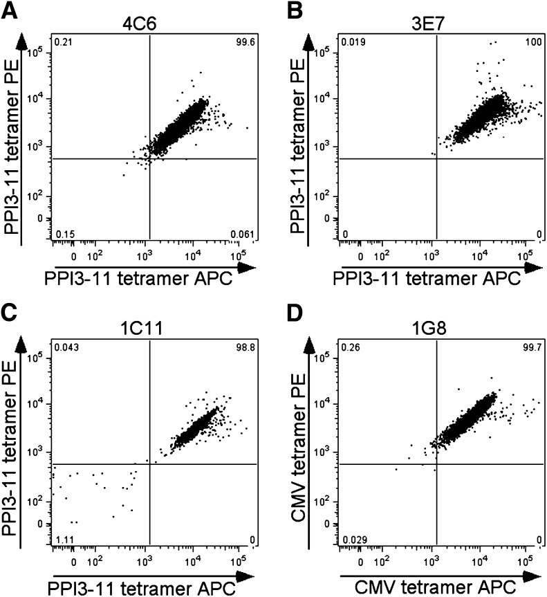
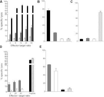
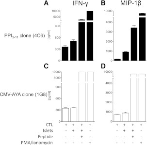
Type 1 diabetes results from T cell-mediated β-cell destruction. The HLA-A*24 class I gene confers significant risk of disease and early onset. We tested the hypothesis that HLA-A24 molecules on islet cells present preproinsulin (PPI) peptide epitopes to CD8 cytotoxic T cells (CTLs). Surrogate β-cell lines secreting proinsulin and expressing HLA-A24 were generated and their peptide ligandome examined by mass spectrometry to discover naturally processed and HLA-A24-presented PPI epitopes. A novel PPI epitope was identified and used to generate HLA-A24 tetramers and examine the frequency of PPI-specific T cells in new-onset HLA-A*24(+) patients and control subjects. We identified a novel naturally processed and HLA-A24-presented PPI signal peptide epitope (PPI(3-11); LWMRLLPLL). HLA-A24 tetramer analysis reveals a significant expansion of PPI(3-11)-specific CD8 T cells in the blood of HLA-A*24(+) recent-onset patients compared with HLA-matched control subjects. Moreover, a patient-derived PPI(3-11)-specific CD8 T-cell clone shows a proinflammatory phenotype and kills surrogate β-cells and human HLA-A*24(+) islet cells in vitro. These results indicate that the type 1 diabetes susceptibility molecule HLA-A24 presents a naturally processed PPI signal peptide epitope. PPI-specific, HLA-A24-restricted CD8 T cells are expanded in patients with recent-onset disease. Human islet cells process and present PPI(3-11), rendering themselves targets for CTL-mediated killing.


Figures

FIG. 1.
FIG. 1.
Generation and characterization of surrogate β-cell line. The myelogenous leukemia cell line K562 is transfected with HLA-A*2402 and INS genes to express PPI (A), such that immunoreactive insulin is detectable in culture supernatants by ELISA, and HLA-A24 (B), shown here detected by flow cytometry using a pan–HLA-ABC monoclonal antibody W6/32. Expression of major histocompatibility complex class I molecules on K562-A24-PPI cells (solid line) was slightly higher than in the K562-A24 cells (dashed line). Isotype control staining was similar in both cell lines; gray filled curve shows K562-A24-PPI isotype staining.
FIG. 2.
FIG. 2.
Distribution of amino acids at all positions including both major anchors (P2 and P9) for all 107 9-mer peptides eluted from HLA-A*24+ K562 cells expressing PPI compared with the amino acid frequency of the human proteome. Data shown as a heatmap in which increased or decreased amino acid frequencies are drawn as a gradient of green or red shades, respectively. Only amino acids that show a significant difference in distribution from the theoretical human proteome are shown (P < 0.05). Heatmap was created using iceLogo software (21). Trp at P2 is highly unusual for HLA-A24 but does not show as significantly upregulated in iceLogo since it is also relatively rare in the human proteome (1.4%). (A high-quality color representation of this figure is available in the online issue.)
FIG. 3.
FIG. 3.
A: Tandem mass spectrometry analysis of collision-induced dissociation revealing the tandem mass spectrum of a peptide of mass (577.86 m/z) in fraction 33. B: The correct identity of the peptide was proven by tandem mass spectrometry of the synthetic compound. C: Amino acid sequence of the peptide with the expected b- and y-fragment ions. Observed fragment ions are underlined. This confirms PPI3–11 (LMWRLLPLL) as naturally processed and presented by HLA-A24 on K562-A24-PPI cells. (A high-quality color representation of this figure is available in the online issue.)
FIG. 4.
FIG. 4.
Representative dual tetramer staining (PE- and APC-labeled) of PBMCs from a patient with type 1 diabetes. Gated, live CD3+CD8 cells are shown stained with PPI3–11-loaded (A) and CMV-AYA–loaded (B) tetramers. C: Percentage of CD8 T cells that are dual tetramer positive when stained with PPI3–11-loaded reagents in 10 patients with recent onset type 1 diabetes and 10 control subjects, all bearing HLA-A*24. Horizontal lines represent medians, and levels are significantly higher in patients than in control subjects. D: Staining with dual tetramers loaded with CMV-AYA. Percentage dual tetramer-positive cells is similar in patients and control subjects.
FIG. 5.
FIG. 5.
AC: Representative staining of three CD8 T-cell clones generated from a single patient that are dual PPI3–11-tetramer positive and a single clone staining with CMV-AYA–loaded reagents (D).
FIG. 6.
FIG. 6.
Cytotoxicity assays using 4C6 PPI3–11- and 1G8 CMV-AYA–specific CD8 T-cell clones. A: 4C6 PPI3–11 CTLs kill K562-A24-PPI cells naturally processing and presenting PPI3–11 (gray bar) at a range of effector-to-target ratios, with increasing killing as the ratio increases. Higher killing is observed, and peaks at a ratio of 12:1, when K562-A24 cells are pulsed with PPI3–11 (black bar). Low-level killing is seen with K562-A24 pulsed with peptide diluent only (open bar). Results are representative of three experiments, and error bars are SEMs from triplicate wells. B: 4C6 PPI3–11 CTLs kill K562-A24 cells pulsed with PPI3–11 peptide (black bar) and K562-A24-PPI naturally presenting PPI3–11 (gray bar), but only background killing of K562-A24 cells pulsed with irrelevant CMV-AYA peptide (cross-hatched bar) or peptide diluent only (open bar) is seen. Effector-to-target ratio 25:1. C: 1G8 CMV-AYA CTLs kill K562-A24 target cells pulsed with CMV-AYA peptide (cross-hatched bar), but show background killing only in the presence of K562-A24 pulsed with PPI3–11 (black bar), K562-A24-PPI naturally presenting PPI (gray bar), or K562-A24 pulsed with peptide diluent only (open bar). Effector-to-target ratio 25:1. D: 4C6 PPI3–11 CTLs kill HLA-A*24+ human islet cells naturally processing and presenting PPI3–11 (gray bar). Only low-level killing is seen with CMV-AYA CTLs (open bar). Both PPI3–11 and CMV-AYA CTLs kill human islet cells to comparable, high levels when targets are pulsed with cognate peptide (black and cross-hatched bar, respectively). Results are a single experiment, and error bars are SEMs from triplicate wells. E: There is robust, high-level killing of human HLA-A*0201+ islets by the previously characterized CTL clone 3F2, specific for PPI15–24 when presented by HLA-A2 (5) either when the islet cells are pulsed with PPI15–24 (gray bar) or when islets are naturally presenting PPI15–24 (open bar). In contrast, killing of the same HLA-A*2+ islets (unpulsed) is only at background levels in the presence of HLA-A24–restricted 4C6 PPI3–11 CTLs (black bar) and HLA-A24 1G8 CMV-AYA CTLs (cross-hatched bar). Effector-to-target ratio 50:1. Experiments represented in B, C, and E were performed in parallel to one another with the same passage of CTL.
FIG. 7.
FIG. 7.
Cytokine production by 4C6 PPI3–11- and 1G8 CMV-AYA–specific CD8 T-cell clones after coculture with HLA-A*24+ human islet cells. 4C6 PPI3–11 CTLs make IFN-γ (A) and MIP-1β (B) in response to culture with HLA-A*24+ human islet cells. Levels are maximal when human islet cells are pulsed with PPI3–11 peptide or CTLs are stimulated with PMA/ionomycin. C and D: 1G8 CMV-AYA CTLs fail to make these cytokines in response to culture with unmanipulated human islet cells but show full cytokine production capability when cells are pulsed with cognate peptide or stimulated with PMA/ionomycin. Results are a single experiment and error bars are SEMs from duplicate wells.

References

    1. Bottazzo GF, Dean BM, McNally JM, MacKay EH, Swift PG, Gamble DR. In situ characterization of autoimmune phenomena and expression of HLA molecules in the pancreas in diabetic insulitis. N Engl J Med 1985;313:353–360 - PubMed
    1. Willcox A, Richardson SJ, Bone AJ, Foulis AK, Morgan NG. Analysis of islet inflammation in human type 1 diabetes. Clin Exp Immunol 2009;155:173–181 - PMC - PubMed
    1. Foulis AK, Farquharson MA, Hardman R. Aberrant expression of class II major histocompatibility complex molecules by B cells and hyperexpression of class I major histocompatibility complex molecules by insulin containing islets in type 1 (insulin-dependent) diabetes mellitus. Diabetologia 1987;30:333–343 - PMC - PubMed
    1. Itoh N, Hanafusa T, Miyazaki A, et al. Mononuclear cell infiltration and its relation to the expression of major histocompatibility complex antigens and adhesion molecules in pancreas biopsy specimens from newly diagnosed insulin-dependent diabetes mellitus patients. J Clin Invest 1993;92:2313–2322 - PMC - PubMed
    1. Skowera A, Ellis RJ, Varela-Calviño R, et al. CTLs are targeted to kill beta cells in patients with type 1 diabetes through recognition of a glucose-regulated preproinsulin epitope [corrected in: J Clin Invest 2009;119:2844]. J Clin Invest 2008;118:3390–3402 - PMC - PubMed

Publication types

MeSH terms