. 2012 Nov 1;520(16):3687-706.
doi: 10.1002/cne.23123.

Systematic mapping of fragile X granules in the mouse brain reveals a potential role for presynaptic FMRP in sensorimotor functions

Affiliations

Systematic mapping of fragile X granules in the mouse brain reveals a potential role for presynaptic FMRP in sensorimotor functions

Michael R Akins et al. J Comp Neurol. .

Abstract

Loss of Fragile X mental retardation protein (FMRP) leads to Fragile X syndrome (FXS), the most common form of inherited intellectual disability and autism. Although the functions of FMRP and its homologs FXR1P and FXR2P are well studied in the somatodendritic domain, recent evidence suggests that this family of RNA binding proteins also plays a role in the axonal and presynaptic compartments. Fragile X granules (FXGs) are morphologically and genetically defined structures containing Fragile X proteins that are expressed axonally and presynaptically in a subset of circuits. To further understand the role of presynaptic Fragile X proteins in the brain, we systematically mapped the FXG distribution in the mouse central nervous system. This analysis revealed both the circuits and the neuronal types that express FXGs. FXGs are enriched in circuits that mediate sensory processing and motor planning-functions that are particularly perturbed in FXS patients. Analysis of FXG expression in the hippocampus suggests that CA3 pyramidal neurons use presynaptic Fragile X proteins to modulate recurrent but not feedforward processing. Neuron-specific FMRP mutants revealed a requirement for neuronal FMRP in the regulation of FXGs. Finally, conditional FMRP ablation demonstrated that FXGs are expressed in axons of thalamic relay nuclei that innervate cortex, but not in axons of thalamic reticular nuclei, striatal nuclei, or cortical neurons that innervate thalamus. Together, these findings support the proposal that dysregulation of axonal and presynaptic Fragile X proteins contribute to the neurological symptoms of FXS.


Conflict of interest statement

Conflict of Interest: The authors declare no competing interests.

Figures

Figure 1
Figure 1
Specificity of anti-FXR2P. BU38 antiserum was generated as described in Methods. (A) BU38 specifically recognized a single polypeptide with an apparent mobility of ~90kD (arrow) in extracts of whole brain from P15 wild type, but not FXR2−/− mice. (B) BU38 does not recognize FMRP or FXR1P. Lysates from COS cells transfected with constructs encoding EGFP alone or EGFP-tagged FXR2P, FMRP or FXR1P were probed with BU38 (‘FXR2P’). This antibody recognized EGFP-FXR2P (arrow) as well as the endogenous FXR2P (asterisks). The expression of all transfected constructs was verified by probing the same samples with anti-GFP (‘GFP’).
Figure 2
Figure 2
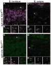
Cellular origin of FXGs. FXG expression in hippocampus of wild type (A–D) and synapsin-Cre fmr1 cKO (E–H) mice. FXGs in wild type mossy fibers in stratum lucidum (A; inset in B) contained both FXR2P (green) and FMRP (magenta). FXGs in wild type commissural/associational fibers in stratum oriens (C; inset in D) also contained both FXR2P and FMRP. In contrast, FXGs in cKO mice, where FMRP is selectively ablated in neurons, contained FXR2P but not FMRP in both stratum lucidum (E; inset in F) and stratum oriens (G; inset in H). Note that FXGs in these populations were also more abundant than in the wild type animals. These observations indicate that FXGs are of neuronal origin. Scale bar in A = 10 μm for A, C, E, G; 2.5 μm in B, D, F, H.
Figure 3
Figure 3
FXGs are expressed in fiber tracts and along axons. (A; inset in B) FXGs (arrows in B) in the striatum localized to neurofilament-expressing fiber tracts, but were absent in the surrounding neuropil. Single confocal plane for A and B. (C; inset in D) FXGs (arrows in D) occurred frequently along neurofilament-expressing axons in neocortex. C is a collapsed z-stack of three optical planes; D is from a single optical plane. NF: neurofilament; FMRP: Fragile X mental retardation protein. Scale bar = 20 μm in A, C; 7 μm in B, 10 μm in D.
Figure 4
Figure 4
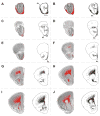
Mapping FXGs in the mouse olfactory bulb. (A) Montage of a wild type P15 olfactory bulb section stained with anti-FXR2P. (B–D) Inset from A showing granules in the olfactory nerve layer. (B) Original image. (C) Image after processing to highlight granules. (D) FXGs were manually annotated (red dots). White arrows in B–D indicate granules identified as FXGs by this methodology; black arrows indicate FXGs that were filtered out in processing. (E) Original image from A inverted and overlaid with FXGs (red) and contours of morphological features (black lines). Note that FXGs are largely restricted to the olfactory nerve layer and glomerular layer. (F) FXGs (black) are overlaid upon a drawing of the section in E. See Table 2 for abbreviations. Scale bar = 500 μm in A, E, F; 25 μm in B–D.
Figure 5
Figure 5
An overview of FXG distribution in the brain. Montage of a saggital section from a P15 wild type brain. (A) FXGs (red) overlaid on an image of the section. (B) FXGs (black) overlaid upon contours. FXGs showed a restricted distribution, with highest expression in olfactory bulb, rostral neocortex, hippocampus, thalamus, and brainstem. Inset: Detailed view of FXG distribution in the thalamus. Note the enriched FXG expression in VA, VL, AV and TRN. See Table 2 for abbreviations. Scale bar = 1 mm in main image; 667 μm in inset.
Figure 6
Figure 6
Systematic mapping of FXG distribution in the P15 mouse brain. Serial coronal sections were immunostained for FXR2P and imaged, processed, annotated and displayed as described in Fig. 4. A single hemisphere from every fifth section is depicted. Each section is shown twice: once with red FXGs overlaid upon an inverted fluorescent image and once with black FXGs overlaid upon a drawing of the contours of the section. Portions lost in tissue preparation are indicated with a dashed line. Inset in Q depicts the ventral hippocampal commissure from a section adjacent to the one mapped in Q. Note the restricted distribution of FXGs to distinct brain regions. FXGs expression was selectively enriched in subregions of several structures including olfactory bulb (A-F), motor cortex (F-P), thalamus (P-Z), hippocampus (Q-Z), and brainstem (Y-PP). Note that FXGs were rare in other brain structures including prefrontal cortex (C-J) and corpus callosum (L-W). See Table 2 for abbreviations. Scale bar for all images = 1 mm.
Figure 6
Figure 6
Systematic mapping of FXG distribution in the P15 mouse brain. Serial coronal sections were immunostained for FXR2P and imaged, processed, annotated and displayed as described in Fig. 4. A single hemisphere from every fifth section is depicted. Each section is shown twice: once with red FXGs overlaid upon an inverted fluorescent image and once with black FXGs overlaid upon a drawing of the contours of the section. Portions lost in tissue preparation are indicated with a dashed line. Inset in Q depicts the ventral hippocampal commissure from a section adjacent to the one mapped in Q. Note the restricted distribution of FXGs to distinct brain regions. FXGs expression was selectively enriched in subregions of several structures including olfactory bulb (A-F), motor cortex (F-P), thalamus (P-Z), hippocampus (Q-Z), and brainstem (Y-PP). Note that FXGs were rare in other brain structures including prefrontal cortex (C-J) and corpus callosum (L-W). See Table 2 for abbreviations. Scale bar for all images = 1 mm.
Figure 7
Figure 7
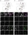
Genetic dissection of FXG origin in thalamocortical circuitry. Boxes in A and G indicate where in the circuit images in B–F and H–L, respectively, were acquired: (B, H) motor cortex, (C, I) external capsule, (D, J,) striatum, (E, K) VAL, and (F, L) TRN. (A) Basic diagram of circuits connecting the thalamus and motor cortex in wild type mice. Corticocortical connections are also indicated. Cortical neurons (red) form synapses onto each other as well as projecting onto thalamic neurons in the ventral anterior and ventral lateral nuclei (VAL; dark blue) and in the reticular nucleus (TRN; light blue). VAL neurons project to both neocortex and TRN. In wild type brain all FXGs in these regions contained both FXR2P (green) and FMRP (magenta). (G) Circuit diagram from the synapsin-Cre cKO mice. All neuronal components are present as in wild type, but FMRP expression was selectively ablated in VAL neurons (cross-hatching). FXGs in striatum (J), VAL (K), and TRN (L) of the cKO mice contained FXR2P but not FMRP (arrowheads). FXGs in motor cortex (H) and subcortical fiber tracts (I) of the cKO mice consisted of two populations: those containing FMRP (arrows) and those lacking FMRP (arrowheads). Scale bar = 10 μm for B–F, H–L.

References

    1. Akins MR, Berk-Rauch HE, Fallon JR. Presynaptic translation: stepping out of the postsynaptic shadow. Front Neural Circuits. 2009;3:17. - PMC - PubMed
    1. Akins MR, Greer CA. Cytoskeletal organization of the developing mouse olfactory nerve layer. J Comp Neurol. 2006;494:358–367. - PMC - PubMed
    1. Baranek GT, Roberts JE, David FJ, Sideris J, Mirrett PL, Hatton DD, Bailey DB., Jr Developmental trajectories and correlates of sensory processing in young boys with fragile X syndrome. Phys Occup Ther Pediatr. 2008;28:79–98. - PubMed
    1. Bassell GJ, Warren ST. Fragile X Syndrome: Loss of Local mRNA Regulation Alters Synaptic Development and Function. Neuron. 2008;60:201–214. - PMC - PubMed
    1. Bontekoe CJM, McIlwain KL, Nieuwenhuizen IM, Yuva-Paylor LA, Nellis A, Willemsen R, Fang Z, Kirkpatrick L, Bakker CE, McAninch R, et al. Knockout mouse model for Fxr2: a model for mental retardation. Hum Mol Genet. 2002;11:487–498. - PubMed

Publication types

Substances