. 2012 Jun 8;287(24):20491-503.
doi: 10.1074/jbc.M112.354761. Epub 2012 Apr 20.

Microphthalmia-associated transcription factor (MITF) promotes differentiation of human retinal pigment epithelium (RPE) by regulating microRNAs-204/211 expression

Affiliations

Microphthalmia-associated transcription factor (MITF) promotes differentiation of human retinal pigment epithelium (RPE) by regulating microRNAs-204/211 expression

Jeffrey Adijanto et al. J Biol Chem. .

Abstract

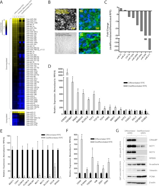
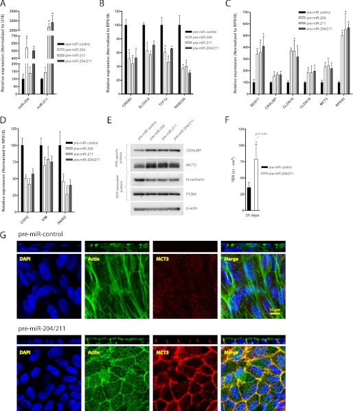
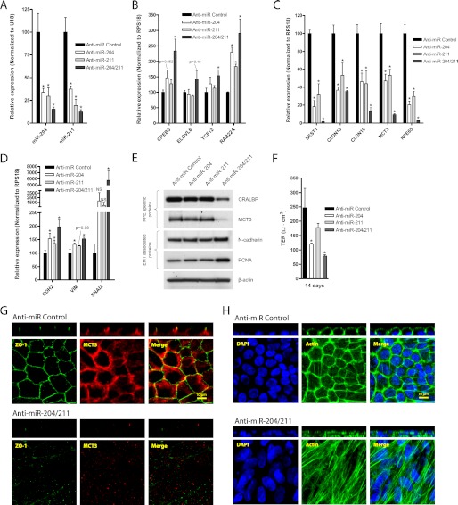
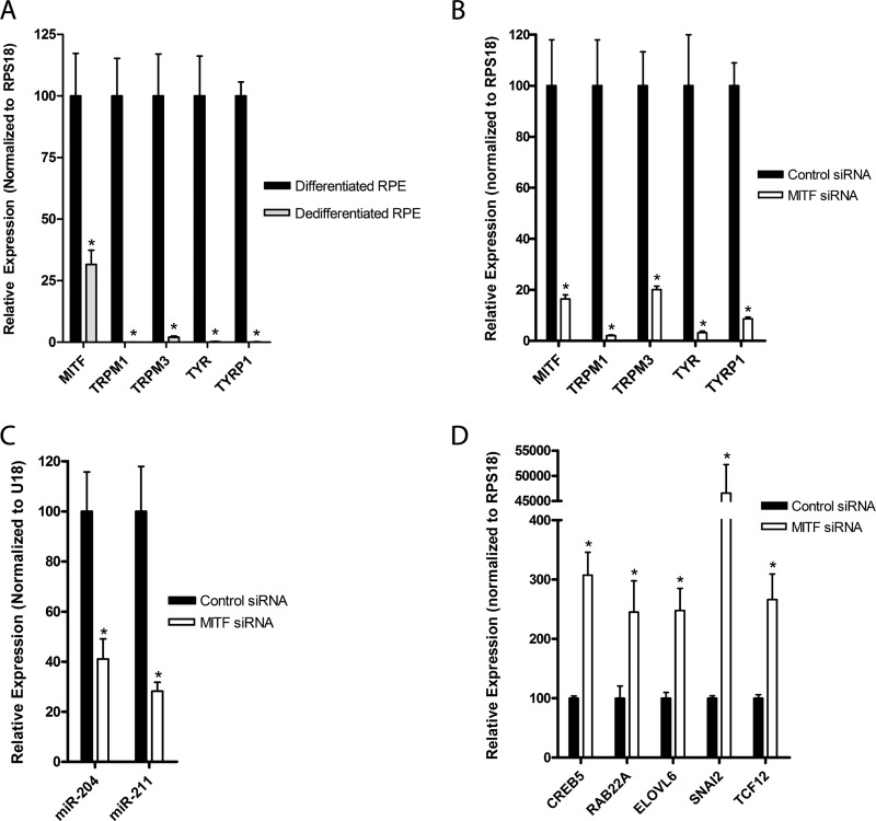
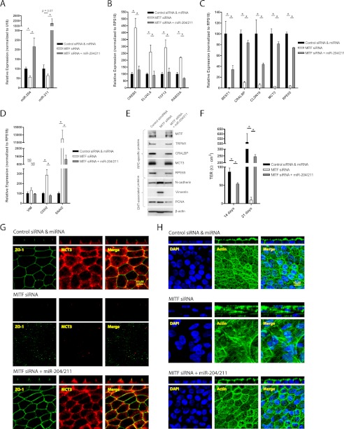
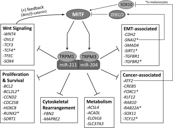
The retinal pigment epithelium (RPE) plays a fundamental role in maintaining visual function and dedifferentiation of RPE contributes to the pathophysiology of several ocular diseases. To identify microRNAs (miRNAs) that may be involved in RPE differentiation, we compared the miRNA expression profiles of differentiated primary human fetal RPE (hfRPE) cells to dedifferentiated hfRPE cells. We found that miR-204/211, the two most highly expressed miRNAs in the RPE, were significantly down-regulated in dedifferentiated hfRPE cells. Importantly, transfection of pre-miR-204/211 into hfRPE cells promoted differentiation whereas adding miR-204/211 inhibitors led to their dedifferentiation. Microphthalmia-associated transcription factor (MITF) is a key regulator of RPE differentiation that was also down-regulated in dedifferentiated hfRPE cells. MITF knockdown decreased miR-204/211 expression and caused hfRPE dedifferentiation. Significantly, co-transfection of MITF siRNA with pre-miR-204/211 rescued RPE phenotype. Collectively, our data show that miR-204/211 promote RPE differentiation, suggesting that miR-204/211-based therapeutics may be effective treatments for diseases that involve RPE dedifferentiation such as proliferative vitreoretinopathy.


Figures

FIGURE 1.
FIGURE 1.
RPE dedifferentiation is characterized by loss of epithelial phenotype, changes in miRNA profile, and significant alterations in mRNA and protein expression. Differentiated hfRPE cells (passage 1 (P1)) were grown on Transwell filters over 4 weeks to obtain a differentiated RPE monolayer. A, dedifferentiated RPE cells that migrated from the free edge of confluent hfRPE monolayer were isolated, and microRNA microarray was performed to compare their miRNA expression levels with that of differentiated RPE cells. B, in a different model, dedifferentiated RPE cells were obtained by passaging P1 hfRPE cells at low cell density twice (P1 to P2 at 1%, P2 to P3 at 30%) on 100-mm culture dishes. C, the expression of several miRNAs that were significantly altered in the microarray analysis was verified using TaqMan microRNA assay. D–F, to compare mRNA expression of differentiated versus dedifferentiated RPE cells, qRT-PCR was performed to evaluate the expression of miR-204/211 targets (D), genes involved in barrier, nutrient/ion transport, and RPE-specific functions (E), and genes that are commonly up-regulated in EMT (F). G, Western blot shows that RPE dedifferentiation involves a loss of RPE-specific proteins and an increase in EMT-associated proteins. Statistically significant changes (p < 0.05) are marked with asterisks. Error bars, S.D.
FIGURE 2.
FIGURE 2.
miR-204/211 promote RPE function and integrity. A–D, hfRPE cells were seeded at 15% density on Transwell filters and transfected (twice; days 0 and 3) with control pre-miRNA (50 nm), pre-miR-204 (25 nm + 25 nm control pre-miRNA), pre-miR-211 (25 nm + 25 nm control pre-miRNA), or pre-miR-204/211 (25 nm each) and cultured for 7 days. In these samples, qRT-PCR was performed to compare relative expression of mature miR-204/211 (A), miR-204/211 targets (B), RPE-specific genes (C), and EMT-associated genes (D). E, in a parallel experiment with identical treatment but grown over 21 days, Western blotting was performed to analyze relative expression of RPE-specific and EMT-associated proteins. F, TER of these samples were measured on the 21st day to evaluate barrier function. G, a set of these samples was fixed and immunostained with DAPI, phalloidin (actin filaments), and MCT3. The confocal vertical (Z-X) sections of the samples are shown in panels above their corresponding en-face (X-Y) representations. Statistically significant changes (p < 0.05) are marked with asterisks. Error bars, S.D.
FIGURE 3.
FIGURE 3.
Inhibition of miR-204/211 results in loss of RPE morphology and phenotype. A–D, hfRPE cells were seeded at 30% cell density on Transwell filters and transfected (twice; days 0 and 3) with control anti-miR (50 nm), anti-miR-204 (25 nm + 25 nm control anti-miR), anti-miR-211 (25 nm + 25 nm control anti-miR), or both anti-miR-204/211 (25 nm each) and cultured for 7 days. In these samples, qRT-PCR was performed to compare relative expression of mature miR-204/211 (A), miR-204/211 targets (B), RPE-specific genes (C), or EMT-associated genes (D). E, in a parallel experiment with identical treatment but grown over 10 days, Western blotting was performed to analyze protein expression RPE-specific and EMT-associated proteins. F, in a separate experiment with the same treatment (two transfections at days 0 and 3) but grown over 14 days, TER was measured with EVOM to evaluate RPE barrier function. G and H, from the experiment in which RPE cells were treated with anti-miRs and grown over 10 days, a set of samples was fixed and immunostained with ZO-1 and MCT3 (G) and DAPI and phalloidin (actin filaments) (H). The confocal vertical (Z-X) sections of the samples are shown in panels above their corresponding en-face (X-Y) representations. Statistically significant changes (p < 0.05) are marked with asterisks. Error bars, S.D.
FIGURE 4.
FIGURE 4.
MITF regulates miR-204/211 expression in RPE. A, qRT-PCR of was performed to compare expression of MITF and its target genes (TRPM1, TRPM3, TYR, TYRP1) in differentiated versus dedifferentiated RPE cells. B–D, hfRPE cells were seeded at 30% density on Transwell filters and transfected twice (days 0 and 3) with control versus MITF siRNA (30 nm each), and qRT-PCR was performed to determine relative expression of MITF and its target genes (B), mature miR-204/211 (C), and miR-204/211 target genes (D). Statistically significant changes (p < 0.05) are marked with asterisks. Error bars, S.D.
FIGURE 5.
FIGURE 5.
MITF knockdown causes loss of miR-204/211 and RPE phenotype that can be prevented by transfection with pre-miR-204/211. A–D, hfRPE cells were seeded at 30% cell density and transfected (twice; days 0 and 3) with control siRNA (30 nm) + control pre-miRNA (30 nm), MITF siRNA (30 nm) + control pre-miRNA (30 nm), or MITF siRNA (30 nm) + pre-miR-204/211 (15 nm each) and cultured for 7 days. In these samples, qRT-PCR was performed to compare relative expression of mature miR-204/211 (A), miR-204/211 targets (B), RPE-specific genes (C), and EMT-associated genes (D). E, in a parallel experiment with identical treatment but grown over 21 days, Western blotting was performed to analyze protein expression RPE-specific and EMT-associated proteins. F–H, in these samples TER was measured with EVOM (14 and 21 days) to evaluate RPE barrier function (F), and a set of these samples was fixed and stained with ZO-1 and MCT3 antibodies (G) and DAPI and phalloidin (actin filaments) (H). The confocal vertical (Z-X) sections of the samples are shown in panels above their corresponding en-face (X-Y) representations. Statistically significant changes (p < 0.05) are marked with asterisks. Error bars, S.D.
FIGURE 6.
FIGURE 6.
miR-204/211 target genes are involved in various cellular functions. miR-204/211 targets were obtained from TargetScan, miRanda, PicTar, and miRDB. These targets were classified into genes that are known to be involved in Wnt signaling, proliferation and survival, cytoskeletal rearrangement, metabolism, cancer, and EMT. Experimentally confirmed miR-204/211 targets are marked with an asterisk.

References

    1. Strauss O. (2005) The retinal pigment epithelium in visual function. Physiol. Rev. 85, 845–881 - PubMed
    1. Adijanto J., Banzon T., Jalickee S., Wang N. S., Miller S. S. (2009) CO2-induced ion and fluid transport in human retinal pigment epithelium. J. Gen. Physiol. 133, 603–622 - PMC - PubMed
    1. Lamb T. D., Pugh E. N., Jr. (2004) Dark adaptation and the retinoid cycle of vision. Prog. Retin. Eye Res. 23, 307–380 - PubMed
    1. Kevany B. M., Palczewski K. (2010) Phagocytosis of retinal rod and cone photoreceptors. Physiology 25, 8–15 - PMC - PubMed
    1. Steele F. R., Chader G. J., Johnson L. V., Tombran-Tink J. (1993) Pigment epithelium-derived factor: neurotrophic activity and identification as a member of the serine protease inhibitor gene family. Proc. Natl. Acad. Sci. U.S.A. 90, 1526–1530 - PMC - PubMed

Publication types

MeSH terms