

Classification and lateralization of temporal lobe epilepsies with and without hippocampal atrophy based on whole-brain automatic MRI segmentation
- PMID: 22523539
- PMCID: PMC3327701
- DOI: 10.1371/journal.pone.0033096
Classification and lateralization of temporal lobe epilepsies with and without hippocampal atrophy based on whole-brain automatic MRI segmentation
Abstract
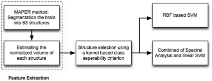
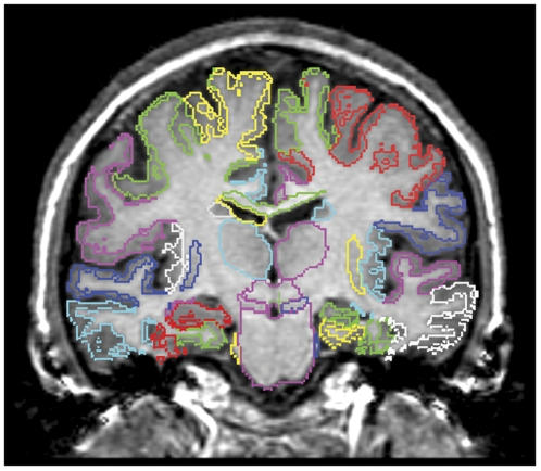
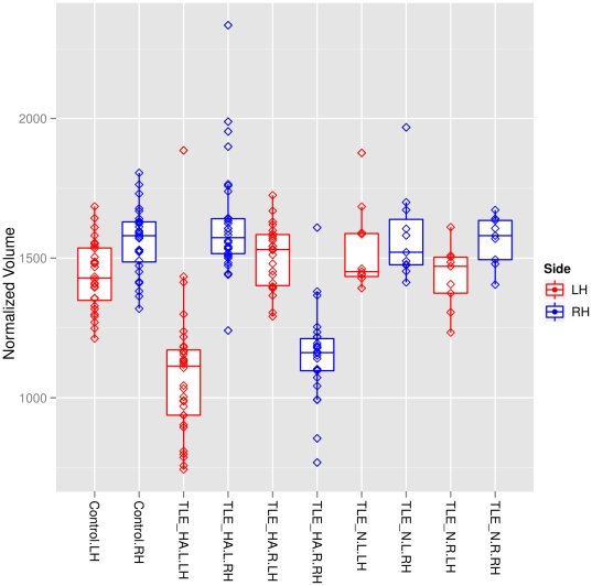
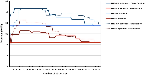
Brain images contain information suitable for automatically sorting subjects into categories such as healthy controls and patients. We sought to identify morphometric criteria for distinguishing controls (n = 28) from patients with unilateral temporal lobe epilepsy (TLE), 60 with and 20 without hippocampal atrophy (TLE-HA and TLE-N, respectively), and for determining the presumed side of seizure onset. The framework employs multi-atlas segmentation to estimate the volumes of 83 brain structures. A kernel-based separability criterion was then used to identify structures whose volumes discriminate between the groups. Next, we applied support vector machines (SVM) to the selected set for classification on the basis of volumes. We also computed pairwise similarities between all subjects and used spectral analysis to convert these into per-subject features. SVM was again applied to these feature data. After training on a subgroup, all TLE-HA patients were correctly distinguished from controls, achieving an accuracy of 96 ± 2% in both classification schemes. For TLE-N patients, the accuracy was 86 ± 2% based on structural volumes and 91 ± 3% using spectral analysis. Structures discriminating between patients and controls were mainly localized ipsilaterally to the presumed seizure focus. For the TLE-HA group, they were mainly in the temporal lobe; for the TLE-N group they included orbitofrontal regions, as well as the ipsilateral substantia nigra. Correct lateralization of the presumed seizure onset zone was achieved using hippocampi and parahippocampal gyri in all TLE-HA patients using either classification scheme; in the TLE-N patients, lateralization was accurate based on structural volumes in 86 ± 4%, and in 94 ± 4% with the spectral analysis approach. Unilateral TLE has imaging features that can be identified automatically, even when they are invisible to human experts. Such morphometric image features may serve as classification and lateralization criteria. The technique also detects unsuspected distinguishing features like the substantia nigra, warranting further study.
Conflict of interest statement
Figures
Comment in
-
Neuroimaging.J Neurol. 2012 Sep;259(9):2009-11. doi: 10.1007/s00415-012-6652-x. J Neurol. 2012. PMID: 22918455 No abstract available.
References
-
- Engel J J. Surgery for seizures. N Engl J Med. 1996;334:647–52. - PubMed
-
- Wiebe S, Blume WT, Girvin JP, Eliasziw M, Effectiveness, et al. A randomized, controlled trial of surgery for temporal-lobe epilepsy. N Engl J Med. 2001;345:311–318. - PubMed
-
- Cascino GD, Jack J C R, Parisi JE, Sharbrough FW, Hirschorn KA, et al. Magnetic resonance imaging-based volume studies in temporal lobe epilepsy: pathological correlations. Ann Neurol. 1991;30:31–6. - PubMed
-
- Paesschen WV, Revesz T, Duncan JS, King MD, Connelly A. Quantitative neuropathology and quantitative magnetic resonance imaging of the hippocampus in temporal lobe epilepsy. Ann Neurol. 1997;42:756–766. - PubMed
-
- Hammers A, Heckemann R, Koepp MJ, Duncan JS, Hajnal JV, et al. Automatic detection and quantification of hippocampal atrophy on MRI in temporal lobe epilepsy: a proof-of-principle study. Neuroimage. 2007;36:38–47. - PubMed
Publication types
MeSH terms
Grants and funding
LinkOut - more resources
Full Text Sources
