. 2012;7(4):e34932.
doi: 10.1371/journal.pone.0034932. Epub 2012 Apr 16.

Ablation of proliferating cells in the CNS exacerbates motor neuron disease caused by mutant superoxide dismutase

Affiliations

Ablation of proliferating cells in the CNS exacerbates motor neuron disease caused by mutant superoxide dismutase

Jean-Nicolas Audet et al. PLoS One. 2012.

Abstract

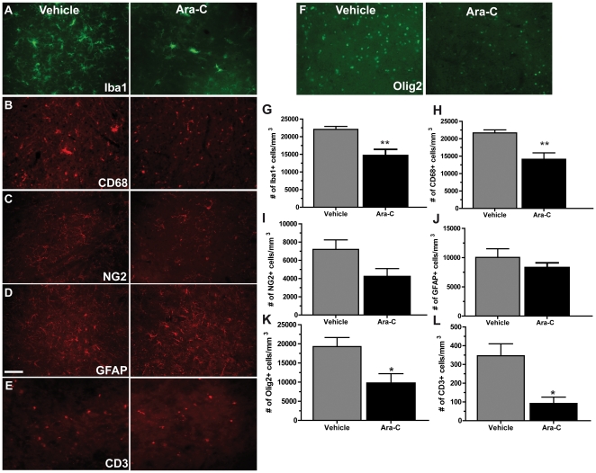
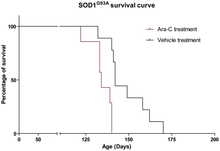
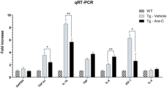
Proliferation of glia and immune cells is a common pathological feature of many neurodegenerative diseases including amyotrophic lateral sclerosis (ALS). Here, to investigate the role of proliferating cells in motor neuron disease, SOD1(G93A) transgenic mice were treated intracerebroventicularly (i.c.v.) with the anti-mitotic drug cytosine arabinoside (Ara-C). I.c.v. delivery of Ara-C accelerated disease progression in SOD1(G93A) mouse model of ALS. Ara-C treatment caused substantial decreases in the number of microglia, NG2+ progenitors, Olig2+ cells and CD3+ T cells in the lumbar spinal cord of symptomatic SOD1(G93A) transgenic mice. Exacerbation of disease was also associated with significant alterations in the expression inflammatory molecules IL-1β, IL-6, TGF-β and the growth factor IGF-1.


Conflict of interest statement

Competing Interests: The authors have declared that no competing interests exist.

Figures

Figure 1
Figure 1. Ara-C treatment caused a decrease in microglia, NG2+ progenitors, oligodendrocytes, astrocytes and T cells in the spinal cord of SOD1G93A mice.
Immunofluorescence for cell markers Iba1 (A), CD68 (B), NG2 (C), GFAP (D), CD3 (E) and Olig2 (F) in the lumbar spinal cord of mutant SOD1 transgenic mice treated with vehicle or Ara-C. (G) Quantification of Iba positive cells showed a 1.5 folds reduction of cells in Ara-C treated mice compared to controls (**p = 0.0086). (H) Quantification of CD68 marker showed a 1.5 folds reduction of cells in Ara-C treated mice compared to controls (**p = 0.0099). (I) Quantification of NG2+ marker showed a 1.7 folds reduction of cells in Ara-C treated mice compared to controls (p = 0.0713) (J) Quantification of GFAP marked cells showed a slightly reduced number of astrocytes (1.2 folds) in Ara-C treated mice compared to controls, although this result was not significant (p = 0.3430). (K) Quantification of Olig2 positive cells showed a 2.0 folds reduction of cells in Ara-C treated mice compared to controls (*p = 0.0236) (L) Quantification of CD3+ cells showed 3.8 folds reduction of cells in Ara-C treated mice compared to controls (*p = 0.0122). All mice were analyzed at 115 days. All values are means ± SEM. Scale bars: 100 µm.
Figure 2
Figure 2. Ablation of proliferating cells in CNS decreases lifespan of SOD1G93A mice.
Kaplan-Meier survival curve shows that transgenic mice Sod1G93A (n = 7) treated with Ara-C between 75 and 115 days had a mean survival of 134 days while untreated SOD1G93A (n = 9) had a mean survival of 141 days. Log-rank test shows that this difference is significant (p = 0.0081).
Figure 3
Figure 3. Motoneuron degeneration of SOD1G93A mice was not affected by Ara-C treatment.
(A) Axons in the ventral root of transgenic mice treated or with Ara-C or vehicle. Quantification of the total number of axons showed no difference between both groups (p = 0.1790). (B) Nissl stain for neuronal cells. Quantification of motoneurons showed no difference between both groups (p = 0.9937). (C) Immunofluorecence for innervation of muscle fibres in the gastrocnemius muscle. Number of innervated, partially innervated and denervated fibre in Ara-C treated mice did not differ from vehicle-treated controls (Denervated: p = 0.8326, partially innervated: p = 0.9281, innervated: p = 0.5504). (A–B) Mice were analyzed at 115 days and (C) end-stage (±140 days). All values are means ± SEM. Scale bars: (A) 100 µm and (B–C) 50 µm.
Figure 4
Figure 4. Modulation of the inflammatory response in Ara-C treated SOD1G93A mice.
Quantitative RT-PCR results (values are normalized to GADPH and relative to wild-type control treated with vehicle; no significant difference was found between wild-type controls treated with vehicle or M-CSF for any marker). Significant differences were found between Ara-C and vehicle treated SOD1G93A in levels of mRNA for TGF-b1 (*p = 0.0148), IL-1b (**p = 0.0072), IL-6 (**p = 0.0014) and IGF-1 (*p = 0.0108). Note that all mRNA levels were significantly higher in vehicle treated transgenic mice compared to WT, treated or not (p<0.0025), except for IL-4 (p = 0.7582). All values are mean ± SEM; n(WT) = 9, n(Tg-Vehicle) = 6, n(Tg-Ara-C) = 15.

References

    1. Clement AM, Nguyen MD, Roberts EA, Garcia ML, Boillee S, et al. Wild-type nonneuronal cells extend survival of SOD1 mutant motor neurons in ALS mice. Science. 2003;302:113–117. - PubMed
    1. Turner BJ, Talbot K. Transgenics, toxicity and therapeutics in rodent models of mutant SOD1-mediated familial ALS. Prog Neurobiol. 2008;85:94–134. - PubMed
    1. Rothstein JD. Current hypotheses for the underlying biology of amyotrophic lateral sclerosis. Ann Neurol. 2009;65(Suppl 1):S3–9. - PubMed
    1. Boillee S, Yamanaka K, Lobsiger CS, Copeland NG, Jenkins NA, et al. Onset and progression in inherited ALS determined by motor neurons and microglia. Science. 2006;312:1389–1392. - PubMed
    1. Yamanaka K, Boillee S, Roberts EA, Garcia ML, McAlonis-Downes M, et al. Mutant SOD1 in cell types other than motor neurons and oligodendrocytes accelerates onset of disease in ALS mice. Proc Natl Acad Sci U S A. 2008;105:7594–7599. - PMC - PubMed

Publication types