. 2012 Apr 19;1(4):350-9.
doi: 10.1016/j.celrep.2012.03.003.

Developmental regulation of chromatin conformation by Hox proteins in Drosophila

Affiliations

Developmental regulation of chromatin conformation by Hox proteins in Drosophila

Marios Agelopoulos et al. Cell Rep. .

Abstract

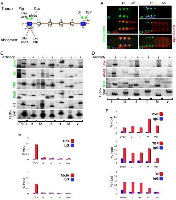
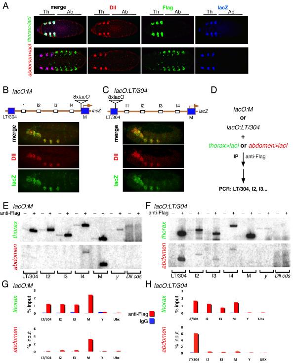
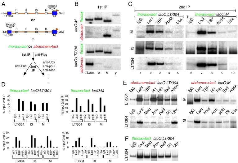
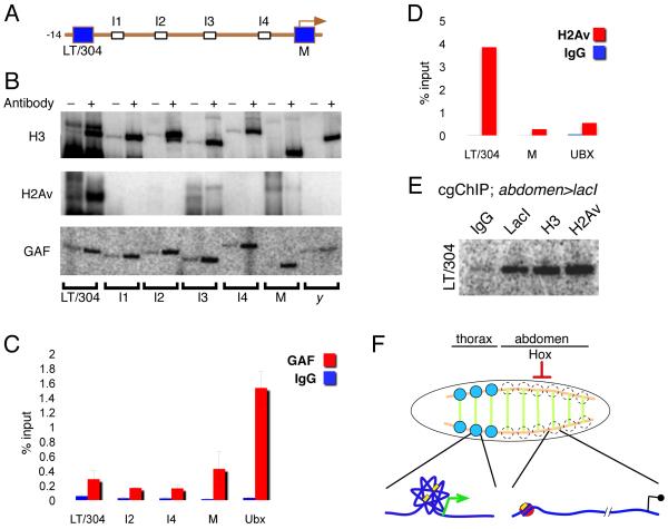
We present a strategy to examine the chromatin conformation of individual loci in specific cell types during Drosophila embryogenesis. Regulatory DNA is tagged with binding sites (lacO) for LacI, which is used to immunoprecipitate the tagged chromatin from specific cell types. We applied this approach to Distalless (Dll), a gene required for limb development in Drosophila. We show that the local chromatin conformation at Dll depends on the cell type: in cells that express Dll, the 5' regulatory region is in close proximity to the Dll promoter. In Dll-nonexpressing cells this DNA is in a more extended configuration. In addition, transcriptional activators and repressors are bound to Dll regulatory DNA in a cell type-specific manner. The pattern of binding by GAGA factor and the variant histone H2Av suggest that they play a role in the regulation of Dll chromatin conformation in expressing and nonexpressing cell types, respectively.


Figures

Figure 1
Figure 1. Whole embryo ChIPs show unique distributions of activators and repressors bound to Dll regulatory regions
(A) Schematic of the 14 kb of DNA 5’ to the start of Dll transcription, showing the positions of the LT/304 CRMs and M element. Positive inputs in the thorax (above the DNA) and negative inputs in the abdomen (below the DNA) are indicated. I1 to I4 are intermediate regions that were monitored by PCR in ChIP experiments. (B) Expression patterns of known Dll activators (Wg, blue; pMad, red) and repressors (Ubx and AbdA, red) relative to Dll expression in the thorax (green). (C) Whole embryo ChIPs using unprogrammed IgG (−) or antibodies (+) to known activators (Arm, Dll, TBP, PolII), Histone3 (H3). IPd chromatin was used as a template for 32P PCRs with the amplicons indicated below the gels. y refers to an amplicon in the yellow gene and serves as a negative control. 12.5 % input shows the signal obtained with input chromatin. (D) Whole embryo ChIPs using unprogrammed IgG (−) or antibodies (+) to a known activator (Mad) or two known repressors (Ubx and AbdA). IPd chromatin was used as a template for 32P PCRs with the amplicons shown below the gels. (E) qPCRs of whole embryo ChIPs comparing the signals obtained with IgG and either anti-Ubx or anti-AbdA for a subset of Dll amplicons. For these repressors, a strong signal was only obtained for LT/304. An amplicon close to the Ubx promoter serves as a negative control. In these and all subsequent qPCRs the error bars represent the standard error of the mean. (F) qPCRs of whole embryo ChIPs comparing the signals obtained with IgG and anti-PolII, anti-TBP, and anti-Dll. For these activators, a strong signal was obtained for LT/304, I2, I4, and M. An amplicon close to the Ubx promoter served as a positive control for PolII and TBP binding, but showed no binding to Dll, as expected.
Figure 2
Figure 2. Cell type specific CRM-promoter interactions at Dll
(A) Ventral views of stage 14 embryos stained for Dll (red), βgal (blue), and Flag-lacI (green). Top row: lacO:M-lacZ; thorax>lacI (thorax-Gal4; UAS-flag-lacI); bottom row: lacO:M-lacZ; abdomen>lacI (abdomen-Gal4; UAS-flag-lacI). The positions of the thoracic and abdominal segments are indicated above the images. Wild type, Dll-like expression of lacZ is observed despite the presence of lacO binding sites and expression of LacI. Note that although abdomen>lacI is expressed in some non-abdominal cells, they do not express Dll (see Methods for more details). (B,C) Lateral views of stage 14 embryos containing the lacO:M (B) and lacO:LT/304 (C) transgenes, stained for Dll (red) and βgal (green). Schematic diagrams of these two lacZ-expressing transgenes are shown above the images. The expression patterns of Dll and lacZ are indistinguishable. (D) Outline of cgChIP experiments for monitoring cell type specific interactions between LT/304 and M using the tools defined in (A-C). (E) 32P PCRs of cgChIPs from lacO:M embryos expressing either thorax>lacI (thorax) or abdomen>lacI (abdomen) as indicated. When Flag-lacI was expressed in the thorax multiple Dll 5’ sequences, but not those from y or the Dll coding sequence (Dll cds), were amplified. In contrast, when Flag-lacI was expressed in the abdomen, only the M element (close to the lacO sites) was amplified. ‘-‘ and ‘+’ above the gels indicate IPs with IgG or anti-Flag, respectively. (F) 32P PCRs of cgChIPs from lacO:LT/304 embryos expressing either thorax>lacI (thorax) or abdomen>lacI (abdomen) as indicated. When Flag-lacI was expressed in the thorax multiple Dll 5’ sequences, but not those from y or the Dll coding sequence (Dll cds), were amplified. In contrast, when Flag-lacI was expressed in the abdomen, only the LT/304 region (close to the lacO sites) was amplified. ‘-‘ and ‘+’ above the gels indicate IPs with IgG or anti-Flag, respectively. (G) qPCR results of cgChIP experiments with lacO:M and thorax>lacI or abdomen>lacI as indicated. The results confirm the 32P PCR results shown in (E). (H) qPCR results for cgChIP experiments with lacO:LT/304 and thorax>lacI or abdomen>lacI as indicated. The results confirm the 32P PCR results shown in (F).
Figure 3
Figure 3. Cell type specific binding of activators and repressors at Dll
(A) Outline of cgChIP experiments for monitoring the presence of factors bound to Dll regulatory regions in thoracic and abdominal cells. (B) 32P PCRs confirming the thoracic-specific interaction between Dll regulatory elements after the primary anti-Flag IP. These data served as a quality control for the primary anti-Flag IP before carrying out any secondary ChIPs as in panels C-F. Independent experiments are shown for both lacO:M and lacO:LT/304. thorax (green) and abdomen (red) refer to thorax>lacI and abdomen>lacI, respectively. (C) 32P PCRs of cgChIPs from thorax>lacI; lacO:LT/304 (left) and thorax>lacI; lacO:M (right) embryos. These PCRs assess the presence of M, I3, and LT/304 sequences following a secondary IP using the antibodies indicated above the gels (IgG, anti-LacI, anti-TBP, anti-AbdA, and anti-Ubx). The results confirm that IPs for LacI and TBP, but not abdominal Hox proteins, pull-down multiple Dll 5’ regions in Dll-expressing cells in the thorax. (D) qPCR measurements of cgChIP experiments for chromatin isolated from thorax>lacI; lacO:LT/304 (left) and thorax>lacI; lacO:M (right). Measurements are for the three Dll sequences (LT/304, I3, and M) after secondary IPs with the antibodies indicated (top gels: IgG, anti-LacI, anti-PolII; bottom gels: IgG, anti-LacI, anti-TBP, anti-AbdA, anti-Ubx). Quantifications are presented as percentages (%) of the qPCR signals obtained from PCRs for the same amplicons after the primary, anti-Flag IP (i.e.; % input 2nd IP). See also Supplementary Figure 1. (E) 32P PCRs of cgChIPs from abdomen>lacI; lacO:LT/304 (left) and abdomen>lacI; lacO:M (right). These PCRs assess the presence of the M or LT/304 sequences following a secondary IP using the antibodies indicated above each gel. IPs for repressors (e.g. Hth, Exd, AbdA, and Ubx) pull-down LT/304 sequences but not M sequences; IPs for activators (TBP, Mad, Arm, PolII, and Dll) fail to pull-down any Dll sequences from abdominal cells. (F) 32P PCRs of cgChIPs from thorax>lacI; lacO:LT/304 embryos. IPs for activators (Mad, Arm, Dll and PolII), but not repressors (Ubx), pull-down these sequences from thoracic cells.
Figure 4
Figure 4. GAF and H2Av have distinct patterns of binding at Dll
(A) Schematic of the −14 kb 5’ Dll regulatory region. (B) Whole embryo ChIPs using anti-H3, anti-H2Av, and anti-GAF as indicated. H2Av, like other repressors, is bound to LT/304, but not other 5’ Dll regions. In contrast, binding of GAF appears to be widespread in the Dll 5’ region. (C) qPCRs of whole embryo ChIPs with anti-GAF, showing widespread binding to the Dll 5’ region, similar to the distribution of other activators (Figure 1). (D) qPCRs of whole embryo ChIPs with anti-H2Av, showing that it is bound to the LT/304 region, but not to the M region. (E) 32P PCR of a cgChIP experiment from abdomen>lacI; lacO:LT/304 embryos, showing that H2Av is bound to the LT/304 region in abdominal cells. (F) Summary of observed cell type specific chromatin configurations in Dll-expressing (thorax) and Dll-non-expressing (abdominal) cells. Thoracic Dll-expressing domains are indicated by the blue circles and occur close to the intersections of Wg expression (green) and Dpp expression (orange). Although Wg and Dpp are present in the same positions in abdominal segments, Dll is repressed in these segments by the abdominal Hox proteins. Our data suggest that in thoracic Dll-expressing cells the entire 5’ region of Dll (with its regulatory elements; yellow boxes) is in a compact state, while in abdominal segments the chromatin structure is more extended and the LT/304 region has H2Av-containing nucleosomes.

References

    1. Agelopoulos M, Thanos D. Epigenetic determination of a cell-specific gene expression program by ATF-2 and the histone variant macroH2A. EMBO J. 2006;25:4843–4853. - PMC - PubMed
    1. Amano T, Sagai T, Tanabe H, Mizushina Y, Nakazawa H, Shiroishi T. Chromosomal dynamics at the Shh locus: limb bud-specific differential regulation of competence and active transcription. Dev Cell. 2009;16:47–57. - PubMed
    1. Apostolou E, Thanos D. Virus Infection Induces NF-kappaB-dependent interchromosomal associations mediating monoallelic IFN-beta gene expression. Cell. 2008;134:85–96. - PubMed
    1. Bartkuhn M, Renkawitz R. Long range chromatin interactions involved in gene regulation. Biochim Biophys Acta. 2008;1783:2161–2166. - PubMed
    1. Biggin MD. Animal transcription networks as highly connected, quantitative continua. Dev Cell. 2011;21:611–626. - PubMed

Publication types

MeSH terms

LinkOut - more resources