Review
. 2012 Apr;54(3):306-16.
doi: 10.1111/j.1440-169X.2012.01342.x.

Interkinetic nuclear migration: a mysterious process in search of a function

Affiliations
Review

Interkinetic nuclear migration: a mysterious process in search of a function

Philip C Spear et al. Dev Growth Differ. 2012 Apr.

Abstract

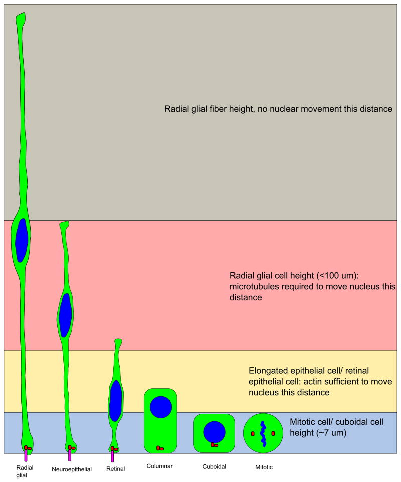
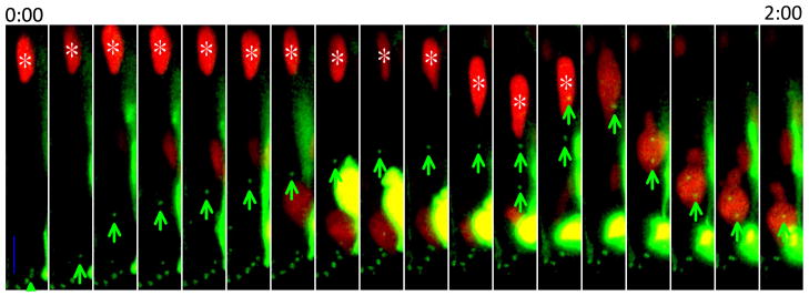
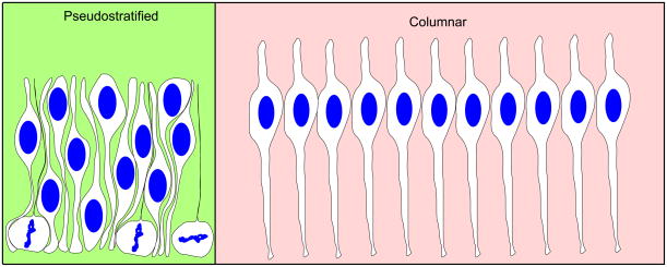
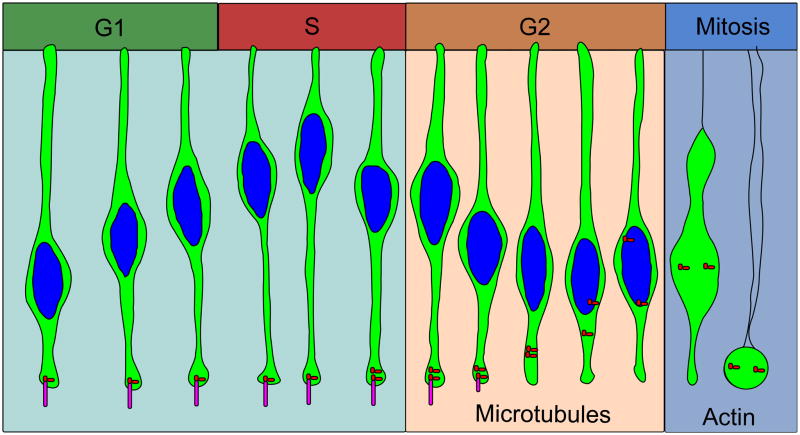
During interkinetic nuclear migration (INM), the nuclei in many epithelial cells migrate between the apical and basal surfaces, coordinating with the cell cycle, and undergoing cytokinesis at the apical surface. INM is observed in a wide variety of tissues and species. Recent advances in time-lapse microscopy have provided clues about the mechanisms and functions of INM. Whether actin or microtubules are responsible for nuclear migration is controversial. How mitosis is initiated during INM is poorly understood, as is the relationship between the cell cycle and nuclear movement. It is possible that the disagreements stem from differences in the tissues being studied, since epithelia undergoing INM vary greatly in terms of cell height and cell fates. In this review we examine the reports addressing the mode and mechanisms that regulate INM and suggest possible functions for this dramatic event.


Figures

Figure 1
Figure 1. Different lengths of epithelial cells may dictate the mechanism of apical nuclear movement for mitosis
Microtubule-dependent and actin-dependent forces may contribute to apical movement during division, but the length of the cell may determine which mechanism contributes the most. Highly elongated cells, such as radial glia, may require the microtubule cytoskeleton. Shorter epithelia undergo shorter nuclear movements to and from the apical surface. In this case, actin-based forces that round up the cell may suffice, with little need for the microtubule cytoskeleton. Cytoplasm of the cells is shown in green, nuclei and DNA are shown in blue. Centrosomes are red and cilia are magenta.
Figure 2
Figure 2. Centrosomes leave the apical surface to initiate mitosis
The intact nucleus is marked with NLS-tdTomato and an asterisk; centrosomes are marked with GFP-centrin and green arrows. The average distance from site of nuclear envelope breakdown to final position during mitosis is 16.5 μm (N = 15). All mitotic cells are observed to undergo apical rounding during mitosis (N = 62). NLS-tdTomato and GFP-centrin were introduced to neural tube cells by in-ovo electroporation at Hamburger/Hamilton stage 12 to 16. Neural tubes were sectioned and imaged less than 24 hours later on an Olympus confocal microscope. Images were taken every 7 minutes. Scale bar = 10 μm
Figure 3
Figure 3. Pseudostratified conformations allow for more cells to be contained with limited surface area
Neural epithelial cells are connected to a limited surface area. Since the nuclei take up more room than the apical endfeet, pseudostratification allows the same number of cells to occupy less space.
Figure 4
Figure 4. Proposed model of INM
Nuclei are moved by actin contraction and along microtubules. Short nuclear movements may be accomplished by actin alone. Longer nuclear movements may require microtubules. Nuclei move basally during G1 phase, reaching a peak distance during S-phase. Nuclei begin moving apically along microtubules using the dynein motor protein during G2 phase. Tpx2 initiates apical nuclear migration at G2 phase. After cilia are lost, centrosomes can move to the nucleus during late G2 to initiate nuclear envelope breakdown and apical rounding, dependent on actin. Cytoplasm of the cells are shown in green, and nuclei are shown in blue. Centrosomes are red dots and cilia are magenta.

References

    1. Afonso C, Henrique D. PAR3 acts as a molecular organizer to define the apical domain of chick neuroepithelial cells. J Cell Sci. 2006;119:4293–304. - PubMed
    1. Ahlstrom JD, Erickson CA. The neural crest epithelial-mesenchymal transition in 4D: a ‘tail’ of multiple non-obligatory cellular mechanisms. Development. 2009;136:1801–1812. - PMC - PubMed
    1. Alvarez-Buylla A, Garci3a-Verdugo JM, Mateo AS, Merchant-Larios H. Primary Neural Precursors and Intermitotic Nuclear Migration in the Ventricular Zone of Adult Canaries. The Journal of Neuroscience. 1998;18:1020–1037. - PMC - PubMed
    1. Austin CP, Feldman DE, Ida JA, Jr, Cepko CL. Vertebrate retinal ganglion cells are selected from competent progenitors by the action of Notch. Development. 1995;121:3637–3650. - PubMed
    1. Basto R, Pines J. The Centrosome Opens the Way to Mitosis. Developmental Cell. 2007;12:475–477. - PubMed

Publication types