. 2012 May 22;51(20):4138-46.
doi: 10.1021/bi300386m. Epub 2012 May 14.

Crystal structures of Xanthomonas campestris OleA reveal features that promote head-to-head condensation of two long-chain fatty acids

Affiliations

Crystal structures of Xanthomonas campestris OleA reveal features that promote head-to-head condensation of two long-chain fatty acids

Brandon R Goblirsch et al. Biochemistry. .

Abstract

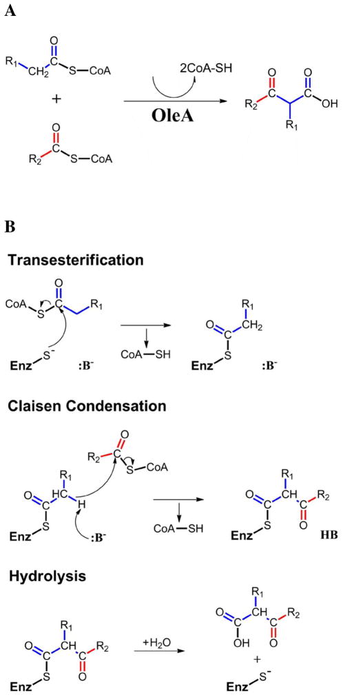
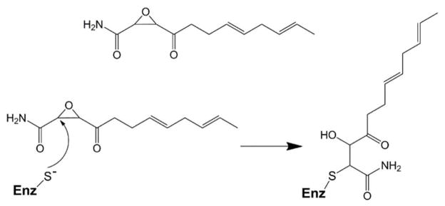
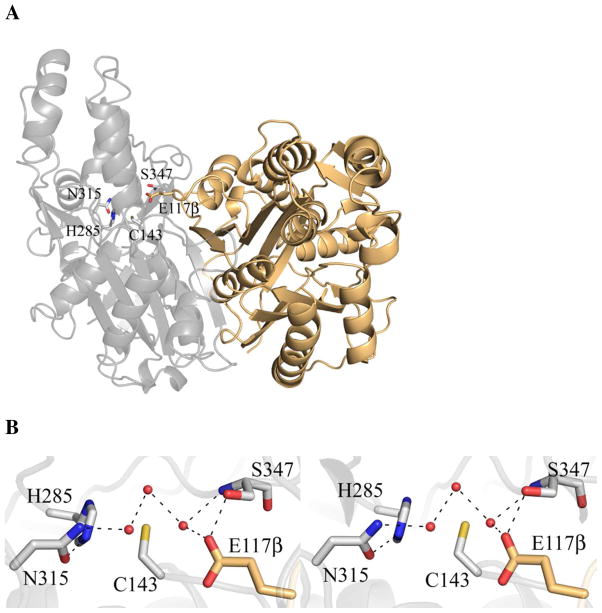
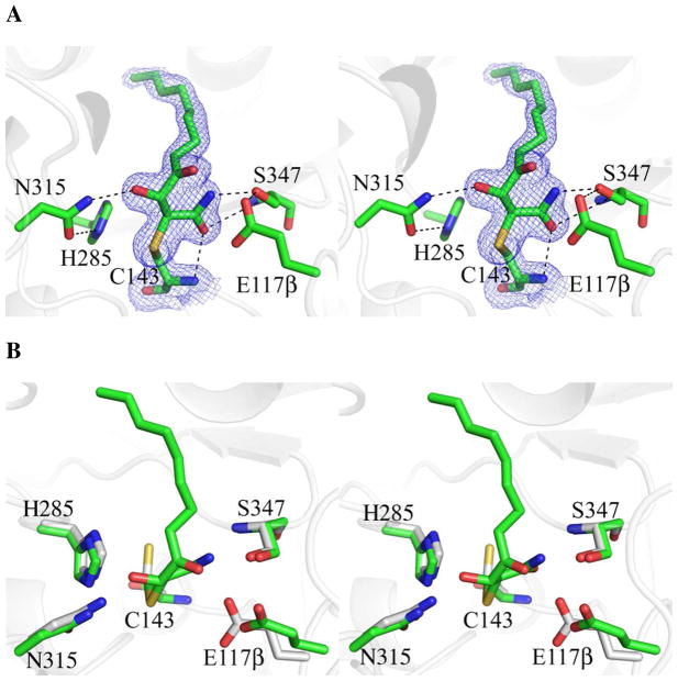
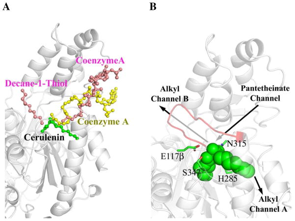
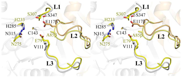
OleA is a thiolase superfamily enzyme that has been shown to catalyze the condensation of two long-chain fatty acyl-coenzyme A (CoA) substrates. The enzyme is part of a larger gene cluster responsible for generating long-chain olefin products, a potential biofuel precursor. In thiolase superfamily enzymes, catalysis is achieved via a ping-pong mechanism. The first substrate forms a covalent intermediate with an active site cysteine that is followed by reaction with the second substrate. For OleA, this conjugation proceeds by a nondecarboxylative Claisen condensation. The OleA from Xanthomonas campestris has been crystallized and its structure determined, along with inhibitor-bound and xenon-derivatized structures, to improve our understanding of substrate positioning in the context of enzyme turnover. OleA is the first characterized thiolase superfamily member that has two long-chain alkyl substrates that need to be bound simultaneously and therefore uniquely requires an additional alkyl binding channel. The location of the fatty acid biosynthesis inhibitor, cerulenin, that possesses an alkyl chain length in the range of known OleA substrates, in conjunction with a single xenon binding site, leads to the putative assignment of this novel alkyl binding channel. Structural overlays between the OleA homologues, 3-hydroxy-3-methylglutaryl-CoA (HMG-CoA) synthase and the fatty acid biosynthesis enzyme FabH, allow assignment of the two remaining channels: one for the thioester-containing pantetheinate arm and the second for the alkyl group of one substrate. A short β-hairpin region is ordered in only one of the crystal forms, and that may suggest open and closed states relevant for substrate binding. Cys143 is the conserved catalytic cysteine within the superfamily, and the site of alkylation by cerulenin. The alkylated structure suggests that a glutamic acid residue (Glu117β) likely promotes Claisen condensation by acting as the catalytic base. Unexpectedly, Glu117β comes from the other monomer of the physiological dimer.


Figures

Figure 1
Figure 1
OleA catalyzed condensation with CoA-charged substrates. (A) The overall reaction. (B) The three steps of the catalytic cycle.
Figure 2
Figure 2
Inhibition of OleA through covalent modification of the active site cysteine by cerulenin.
Figure 3
Figure 3
OleA dimer and active site. (A) The physiological dimer of OleA. The two monomers are drawn in gray and tan cartoon. Each monomer contains one active site. The gray cartoon active site residues are drawn in stick. Note that E117β derives from the neighboring monomer. (B) Stereo view of the OleA active site. The 2Fo-Fc electron density map is contoured to 1.0 σ. Ordered solvent molecules are represented by red spheres. This figure was produced using PyMOL (http://www.pymol.org/).
Figure 4
Figure 4
Stereo view of the OleA active site with cerulenin bound to Cys143. Active site residues and inhibitor are drawn in stick. Solvent molecules are represented by red spheres. Hydrogen bond contacts are indicated by dashed lines. Blue mesh illustrates the 2Fo-Fc electron density maps contoured at 1.0 σ. (A) Active site of OleA co-crystallized with cerulenin (carbon green). (B) Overlay of OleA active sites in the unbound (carbon gray), and cerulenin bound states. The overlay was generated in PyMOL using the program SUPER. This figure was produced using PyMOL (http://www.pymol.org/).
Figure 5
Figure 5
Proposed OleA channel A. (A) The position of the cerulenin alkyl chain and xenon (Xe) atom are consistent with a continuous alkyl binding channel. The cerulenin bound to Cys143 and the xenon are drawn in space-filling colored by atom (carbon, green; Xe, pale yellow). Electron density for the Xe is included (2Fo-Fc contoured at 1.0 σ, blue mesh; anomalous contoured at 9.0 σ, pink mesh). The OleA secondary structural elements that form the channel and contact the cerulenin and Xe are represented in cartoon and colored orange. The bulky hydrophobic side chains that line the most distal part of the channel from Cys143 are also colored orange and drawn in stick. (B) Two helices that line channel A of OleA (gray cartoon) are more closely packed in FabF (PDB ID 1B3N, carbon cyan), FabB (PDB ID 1FJ8, carbon brown), and FabH (PDB ID 2QX1, carbon pink). This figure was produced using PyMOL (http://www.pymol.org/).
Figure 6
Figure 6
Illustration of OleA binding channels. (A) OleA bound with cerulenin overlaid with FabH (bound with decane-1-thiol and coenzyme A, PDB ID 2QX1) and HMG-CoA synthase (bound with coenzyme A, PDB ID 1TXT). OleA is drawn in gray cartoon and cerulenin as a green ball-and-stick model. For clarity, the monomers of FabH and HMG-CoA synthase are omitted and their bound ligands are drawn in ball-and-stick (pink and yellow respectively). Decane-1-thiol occupies the alkyl channel B in FabH. Coenzyme A occupies the pentetheinate channel in both FabH and HMG-CoA synthase. (B) Putative assignment of OleA binding channels. Active site residues are drawn in stick (carbon green) and bound cerulenin drawn in green space-filling. Labeled arrows indicate the position of the alkyl and pantetheinate channels in the OleA monomer (gray cartoon). An eleven residue (239-249) β-hairpin that could only be modeled in one monomer of the P212121 crystal form is drawn as red cartoon. This figure was produced using PyMOL (http://www.pymol.org/).
Figure 7
Figure 7
Overlay of HMG-CoA synthase (PDB ID 1TXT) onto OleA. The dimer of OleA is shown in faded cartoon colored as in Figure 3. Three loop elements (L1, L2, and L3) conserved between OleA and HMG-CoA synthase are highlighted, with L2 originating from the neighboring monomer. Residues forming a single active site in each enzyme are drawn in stick. For simplicity, a single active site cysteine (C143) from OleA is drawn. Residues from OleA are colored by monomer and labeled in black font. The E117β (carbon tan) derives from the L2 loop element making hydrogen bonding contacts with the S347 found in the L1 loop. Spatially equivalent residues from HMG-CoA are labeled in yellow font and colored by monomer. The active site of HMG-CoA synthase is formed completely from one monomer (carbon yellow) including the E79 catalytic base that derives from the L3 loop. In HMG-CoA synthase, the position of the E117β aligns with A85β (carbon pale yellow). The overlay was generated in PyMOL using the program SUPER. This figure was produced using PyMOL (http://www.pymol.org/).

References

    1. Li M, Nørgaard H, Warui DM, Booker SJ, Krebs C, Bollinger JM., Jr Conversion of fatty aldehydes to alka(e)nes and formate by a cyanobacterial aldehyde decarbonylase: cryptic redox by an unusual dimetal oxygenase. J Am Chem Soc. 2011;27:6158–6161. - PMC - PubMed
    1. Lee SK, Chou H, Ham TS, Lee TS, Keasling JD. Metabolic engineering of microorganisms for biofuels production: from bugs to synthetic biology to fuels. Curr Opin Biotechnol. 2008;19:556–563. - PubMed
    1. Sukovich DJ, Seffernick JL, Richman JE, Gralnick JA, Wackett LP. Widespread head-to-head hydrocarbon biosynthesis in bacteria and role of OleA. Appl Environ Microbiol. 2010;76:3850–3862. - PMC - PubMed
    1. Wackett LP. Microbial-based motor fuels: science and technology. Microb Biotechnol. 2008;1:211–225. - PMC - PubMed
    1. Albro PW, Dittmer JC. The biochemistry of long-chain, nonisoprenoid hydrocarbons. IV. Characteristics of synthesis by a cell-free preparation of Sarcina lutea. Biochemistry. 1969;8:3317–3324. - PubMed

Publication types

LinkOut - more resources