. 2012 Jul 15;356(4):209-14.
doi: 10.1016/j.carres.2012.03.028. Epub 2012 Apr 2.

Catalytic mechanism of human UDP-glucose 6-dehydrogenase: in situ proton NMR studies reveal that the C-5 hydrogen of UDP-glucose is not exchanged with bulk water during the enzymatic reaction

Affiliations

Catalytic mechanism of human UDP-glucose 6-dehydrogenase: in situ proton NMR studies reveal that the C-5 hydrogen of UDP-glucose is not exchanged with bulk water during the enzymatic reaction

Thomas Eixelsberger et al. Carbohydr Res. .

Abstract

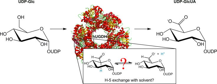
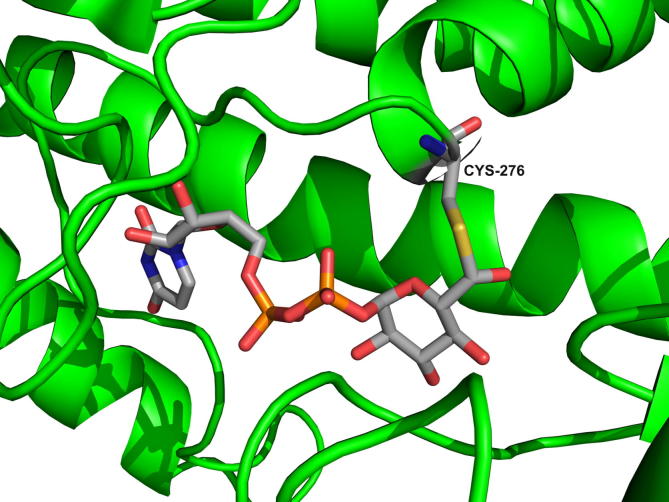
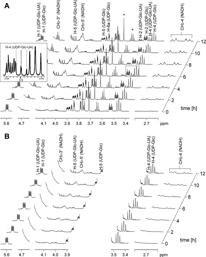
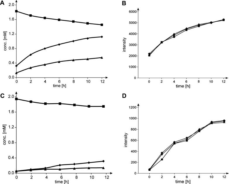
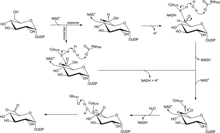
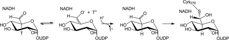
Human UDP-glucose 6-dehydrogenase (hUGDH) catalyzes the biosynthetic oxidation of UDP-glucose into UDP-glucuronic acid. The catalytic reaction proceeds in two NAD(+)-dependent steps via covalent thiohemiacetal and thioester enzyme intermediates. Formation of the thiohemiacetal adduct occurs through attack of Cys(276) on C-6 of the UDP-gluco-hexodialdose produced in the first oxidation step. Because previous studies of the related enzyme from bovine liver had suggested loss of the C-5 hydrogen from UDP-gluco-hexodialdose due to keto-enol tautomerism, we examined incorporation of solvent deuterium into product(s) of UDP-glucose oxidation by hUGDH. We used wild-type enzyme and a slow-reacting Glu(161)→Gln mutant that accumulates the thioester adduct at steady state. In situ proton NMR measurements showed that UDP-glucuronic acid was the sole detectable product of both enzymatic transformations. The product contained no deuterium at C-5 within the detection limit (≤2%). The results are consistent with the proposed mechanistic idea for hUGDH that incipient UDP-gluco-hexodialdose is immediately trapped by thiohemiacetal adduct formation.


Figures

None
Graphical abstract
Figure 1
Figure 1
Close-up representation of the active site of E161Q in a trapped thiohemiacetal enzyme intermediate. Cys276 is the site of covalent modification. The PDB code of the structure is 3KHU.
Figure 2
Figure 2
In situ NMR spectroscopic measurement of hUGDH catalyzed oxidation of UDP-Glc to UDP-GlcUA. The data are presented as stack plot of seven selected spectra in regular intervals of 2 h. Indicative signals of NADH, UDP-Glc, and UDP-Glc-UA are marked and assigned according to literature. Signals of impurities (TRIS from enzyme preparations, ethanol from commercial NAD+) are indicated with asterisks. No signals indicating presence of UDP-gluco-hexodialdose could be detected. Panels A and B show the reaction of wild-type hUGDH and E161Q, respectively. The spectral region from 3.89 ppm to 3.52 ppm is not shown in panel B due to presence of larger signals from impurities (glycerol). In panel A, the area of the spectrum showing the 1H NMR signal of proton H-4 in UDP-GlcUA is also presented enlarged in a box.
Figure 3
Figure 3
Time courses of enzymatic oxidations of UDP-Glc, and determination of the associated change of relative H-5 signal intensity in UDP-GlcUA product. Time courses are shown in panels A (wild-type) and C (E161Q), the symbols used are square (UDP-Glc), triangle (UDP-GlcUA), and diamond (NADH). The low conversion can be explained by a large solvent isotope effect on kcat (see text). Two equivalents of NADH are formed per equivalent UDP-Glc utilized, as expected. Proton signal intensities in UDP-GlcUA are shown in panels B and D (square: H-1; triangle H-4; diamond: H-5) for reactions with wild-type enzyme and E161Q mutant, respectively. In case of deuterium incorporation at C-5, one would see a decrease of the respective proton signal compared to H-1 and H-4.
Figure 4
Figure 4
pH dependence of conversion of UDP-Glc by wild-type hUGDH and E161Q mutant. The symbols are filled circle (wild-type enzyme) and empty circle (mutant). The reaction time was 16 h at 25 °C.
Scheme 1
Scheme 1
Proposed course of the catalytic reaction of UGDH. For clarity reasons, only the herein relevant amino acids of the active site of hUGDH are shown. The concerted mechanism would be inconsistent with exchange of the proton at glucosyl C-5 with solvent. The stepwise mechanism might allow proton exchange due to accumulation of enzyme-bound UDP-gluco-hexodialdose. Unless UDP-gluco-hexodialdose was released from the active site (which probably does not happen as shown later), exchange reaction would have to take place at the active site of the enzyme.
Scheme 2
Scheme 2
Enolization and proton exchange at C-5 of UDP-gluco-hexodialdose. If the aldehyde enolized prior to covalent intermediate formation, tritium label at C-5 could be replaced by hydrogen through label ‘wash-out’ to solvent. Similarly, deuterium could be incorporated (‘washed in’) if the reaction was performed in D2O.

References

    1. Clarkin C.E., Allen S., Kuiper N.J., Wheeler B.T., Wheeler-Jones C.P., Pitsillides A.A. J. Cell. Physiol. 2011;226:749–761. - PubMed
    1. Egger S., Chaikuad A., Kavanagh K.L., Oppermann U., Nidetzky B. Biochem. Soc. Trans. 2010;38:1378–1385. - PubMed
    1. Campbell R.E., Mosimann S.C., van De Rijn I., Tanner M.E., Strynadka N.C. Biochemistry. 2000;39:7012–7023. - PubMed
    1. Egger S., Chaikuad A., Kavanagh K.L., Oppermann U., Nidetzky B. J. Biol. Chem. 2011;286:23877–23887. - PMC - PubMed
    1. Rocha J., Popescu A.O., Borges P., Mil-Homens D., Moreira L.M., Sa´-Correia I., Fialho A.M., Frazão C. J. Bacteriol. 2011;193:3978–3987. - PMC - PubMed

Publication types

MeSH terms

LinkOut - more resources