Review
. 2012 Nov 27;91(21-22):986-91.
doi: 10.1016/j.lfs.2012.04.010. Epub 2012 Apr 13.

Critical metabolic roles of β-cell M3 muscarinic acetylcholine receptors

Affiliations
Review

Critical metabolic roles of β-cell M3 muscarinic acetylcholine receptors

Inigo Ruiz de Azua et al. Life Sci. .

Abstract

Muscarinic acetylcholine (ACh) receptors (mAChRs; M(1)-M(5)) regulate the activity of an extraordinarily large number of important physiological processes. We and others previously demonstrated that pancreatic β-cells are endowed with M(3) mAChRs which are linked to G proteins of the G(q) family. The activation of these receptors by ACh or other muscarinic agonists leads to the augmentation of glucose-induced insulin release via multiple mechanisms. Interestingly, in humans, ACh acting on human β-cell mAChRs is released from adjacent α-cells which express both choline acetyltransferase (ChAT) and the vesicular acetylcholine transporter (vAChT), indicative of the presence of a non-neuronal cholinergic system in human pancreatic islets. In order to shed light on the physiological roles of β-cell M(3) receptors, we recently generated and analyzed various mutant mouse models. Specifically, we carried out studies with mice which overexpressed M(3) receptors or mutant M(3) receptors in pancreatic β-cells or which selectively lacked M(3) receptors or M(3)-receptor-associated proteins in pancreatic β-cells. Our findings indicate that β-cell M(3) receptors play a key role in maintaining proper insulin release and whole body glucose homeostasis and that strategies aimed at enhancing signaling through β-cell M(3) receptors may prove useful to improve β-cell function for the treatment of type 2 diabetes (T2D).


Figures

Fig. 1
Fig. 1
Scheme summarizing the key phenotypes displayed by β-M3-KO and β-M3-Tg mice. See text for details (Gautam et al., 2006).
Fig. 2
Fig. 2
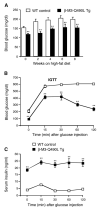
M3-Q490L Tg mice are protected against the detriment metabolic effects associated with the consumption of a high-fat diet. All studies were carried out with male mice maintained on a high-fat diet (n=6 or 7 per group). (A) Blood glucose levels of freely fed β-M3-Q490L Tg mice and WT littermates. (B) I.p. glucose tolerance test (IGTT). Blood glucose levels were measured at the indicated time points following i.p. administration of glucose (2 mg/g body weight). (C) Serum insulin levels following i.p. administration of glucose (2 mg/g body weight). Data are expressed as means± SEM. **P<0.01, as compared to the corresponding WT value. Data were taken from Gautam et al. (2010).
Fig. 3
Fig. 3
Examples of ACh-insensitive mutant M3 mAChRs that can be activated by clozapine-N-oxide (CNO). Both the R-q and R-s mutant receptors contain the Y148C and A238G point mutations (rat M3 receptor sequence). These two point mutations prevent binding of the neurotransmitter ACh (Armbruster et al., 2007; Guettier et al., 2009). However, both R-q and R-s can be activated by CNO with high potency and efficacy (Armbruster et al., 2007; Guettier et al., 2009). CNO is a pharmacologically inert metabolite of clozapine. The two mutant receptors differ in their G protein-coupling properties (R-q: Gq-type G proteins; R-s: Gs; Guettier et al., 2009). Figure was taken from Guettier et al. (2009).
Fig. 4
Fig. 4
RGS4 deficiency results in the selective enhancement of M3 receptor-mediated increases in insulin secretion. Insulin release experiments were carried out with isolated pancreatic islets prepared from WT and RGS4-deficient mice, as described by Ruiz de Azua et al. (2010). Islets were incubated in the presence of a stimulatory concentration of glucose (16.7 mM) in the absence of ligands or in the presence of oxotremorine-M (OXO-M, 0.5 μM), glucagon-like peptide 1 (GLP-1, 10 nM), ADP (100 μM), or arginine vasopressin (AVP, 100 nM). These ligands are known to promote insulin secretion via activation of specific β-cell GPCRs. OXO-M stimulates the release of insulin from pancreatic β-cells via activation of M3 mAChRs (Duttaroy et al., 2004; Zawalich et al., 2004). Data are expressed as means±SEM of three independent experiments, each carried out in triplicate. *P<0.05, ***P<0.001, as compared to the corresponding WT value. Data were taken from Ruiz de Azua et al. (2010).

References

    1. Abramow-Newerly M, Roy AA, Nunn C, Chidiac P. RGS proteins have a signalling complex: interactions between RGS proteins and GPCRs, effectors, and auxiliary proteins. Cell Signal. 2006;18:579–91. - PubMed
    1. Ahrén B. Autonomic regulation of islet hormone secretion—implications for health and disease. Diabetologia. 2000;43:393–410. - PubMed
    1. Ahrén B. Islet G protein-coupled receptors as potential targets for treatment of type 2 diabetes. Nat Rev Drug Discov. 2009;8:369–85. - PubMed
    1. Armbruster BN, Li X, Pausch MH, Herlitze S, Roth BL. Evolving the lock to fit the key to create a family of G protein-coupled receptors potently activated by an inert ligand. Proc Natl Acad Sci U S A. 2007;104:5163–8. - PMC - PubMed
    1. Baggio LL, Drucker DJ. Biology of incretins: GLP-1 and GIP. Gastroenterology. 2007;132:2131–57. - PubMed

Publication types