. 2012 Jun 15;72(12):2931-6.
doi: 10.1158/0008-5472.CAN-11-4097. Epub 2012 Apr 23.

Mice expressing activated PI3K rapidly develop advanced colon cancer

Affiliations

Mice expressing activated PI3K rapidly develop advanced colon cancer

Alyssa A Leystra et al. Cancer Res. .

Abstract

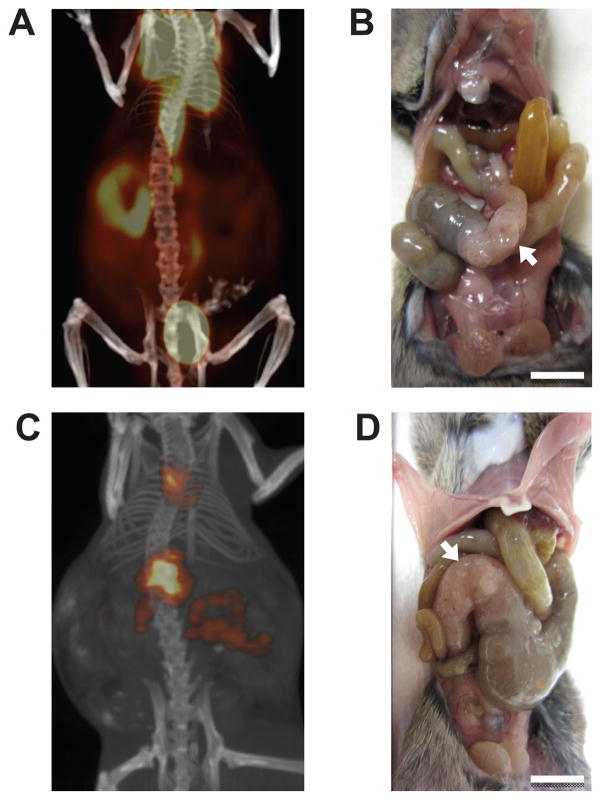
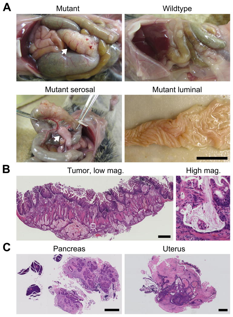
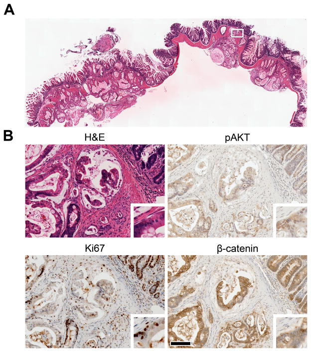
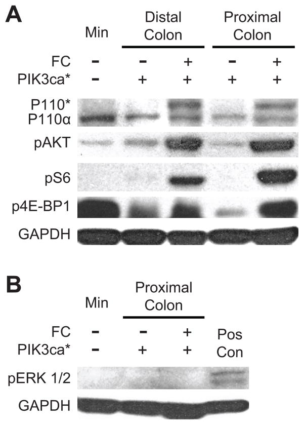
Aberrations in the phosphoinositide 3-kinase (PI3K) signaling pathway play a key role in the pathogenesis of numerous cancers by altering cellular growth, metabolism, proliferation, and apoptosis. Mutations in the catalytic domain of PI3K that generate a dominantly active kinase are commonly found in human colorectal cancers and have been thought to drive tumor progression but not initiation. However, the effects of constitutively activated PI3K upon the intestinal mucosa have not been previously studied in animal models. Here, we show that the expression of a dominantly active form of the PI3K protein in the mouse intestine results in hyperplasia and advanced neoplasia. Mice expressing constitutively active PI3K in the epithelial cells of the distal small bowel and colon rapidly developed invasive adenocarcinomas in the colon that spread into the mesentery and adjacent organs. The histologic characteristics of these tumors were strikingly similar to invasive mucinous colon cancers in humans. Interestingly, these tumors formed without a benign polypoid intermediary, consistent with the lack of aberrant WNT signaling observed. Together, our findings indicate a noncanonical mechanism of colon tumor initiation that is mediated through activation of PI3K. This unique model has the potential to further our understanding of human disease and facilitate the development of therapeutics through pharmacologic screening and biomarker identification.


Conflict of interest statement

Author Information The authors declare competing financial interests: Dr. Jamey Weichert is the founder of Cellectar, Inc. (Madison, WI), which holds the licensing rights to the CLR1404 technology, and therefore has a financial interest in this agent.

Figures

Figure 1
Figure 1. FC+ PIK3ca*+ mice develop intestinal obstruction due to large proximal colon tumors
The tumors exhibit a high avidity for FDG (A) and CLR1404 (C). The mouse was injected with the imaging agent, scanned, and the data was reconstructed using standard algorithms. Following scan acquisition, each mouse was sacrificed and the abdominal wall was dissected revealing tumors in the proximal colon (B and D, arrows). Scale bars: 1 cm.
Figure 2
Figure 2. The large tumors in the proximal colon are invasive mucinous adenocarcinomas
A FC+ PIK3ca*+ mouse and its wild-type littermate were sacrificed at 56 days of age and dissected (A upper left and right, respectively). In the mutant, severe dilation of the small bowel and cecum was due to obstruction by a massive tumor (A upper left, arrow). The tumor was associated with large penetrating blood vessels and thickened mesentery (A lower left, arrow). The intestine was removed, split lengthwise, and splayed out. The epithelium was hyperplastic but no obvious polypoid intermediary was observed (A lower right). The tumor was removed, embedded in paraffin, and cut. Sections were stained with H&E (B). Islands of malignant glands in mucin lakes were evident at higher magnification (B right). One cecal tumor appeared to directly extend to surrounding organs including the pancreas and in the uterus (C). Scale bars: 1 cm (A) and 1 mm (B, and C).
Figure 3
Figure 3. PIK3ca*inducescellular proliferation
A section of a tumor from a 61-day-old FC+ PIK3ca*+ mouse was stained with H&E (A). The tumor invaded through the musculature into the adipose tissue of the colonic mesenteries. The boxed region in A is shown at higher magnification (B). The activation of AKT, a downstream target of PI3K, is revealed by immunohistochemistry (IHC) using antibodies specific for pAKT (B upper right). The majority of nuclei are positive (brown). This change was coupled with an increase in cellular proliferation as measured by IHC using antibodies against Ki67 (B lower left). The invasive adenocarcinomas that develop in this model do not appear to rely on aberrant WNT signaling, as β-catenin is membrane-bound rather than nuclear, as shown by IHC (B lower right). This localization of β-catenin was observed in 10 out of 10 tumors. Insets were taken from the left edge of each panel and magnified two-fold (B). Scale bar: 100 μm.
Figure 4
Figure 4. Activation of the PI3K/AKT/mTOR pathway occurs in the colonic mucosa and tumors of FC+ PIK3ca*+ mice without increasing pERK 1/2
Protein extracts were prepared from scrapings of the mucosa that were taken from either the proximal or distal colon of experimental and control mice. Note that the scrapings from the proximal colon of FC+ PIK3ca*+ mice contained tumor tissue. Protein extracts were also prepared from a colon tumor that was taken from a Min (ApcMin/+) mouse for comparison. p110* is identifiable by western blot in the proximal and distal colon of FC+ PIK3ca*+ mice (A). The level of p110α is similar in all tissues tested. Up-regulation of the PI3K/AKT/mTOR pathway is noted by increased phosphorylation in AKT, S6, and 4E-BP1. Phosphorylation of ERK 1/2 was undetectable in protein extracts from proximal colon tumors of four FC+ PIK3ca*+ mice, a representative is shown (B). DLD-1, a human colorectal cancer cell line that express mutant KRAS, was used as a positive control (Pos Con); GAPDH was used as a loading control.

References

    1. Vivanco I, Sawyers CL. The phosphatidylinositol 3-Kinase AKT pathway in human cancer. Nat Rev Cancer. 2002;2:489–501. - PubMed
    1. Huang CH, Mandelker D, Gabelli SB, Amzel LM. Insights into the oncogenic effects of PIK3CA mutations from the structure of p110alpha/p85alpha. Cell Cycle. 2008;7:1151–1156. - PMC - PubMed
    1. Samuels Y, et al. High frequency of mutations of the PIK3CA gene in human cancers. Science. 2004;304:554. - PubMed
    1. Zhao L, et al. Hot-spot mutations in p110α of phosphatidylinositol 3-kinase (PI3K) Cell Cycle. 2010;9:596–600. - PMC - PubMed
    1. Clapper ML, Cooper HS, Chang WC. Dextran sulfate sodium-induced colitis-associated neoplasia: a promising model for the development of chemopreventive interventions. Acta Pharmacol Sin. 2007;28:1450–1459. - PubMed

Publication types

MeSH terms

Substances