. 2012 May;68(Pt 5):592-600.
doi: 10.1107/S0907444912006749. Epub 2012 Apr 17.

In situ macromolecular crystallography using microbeams

Affiliations

In situ macromolecular crystallography using microbeams

Danny Axford et al. Acta Crystallogr D Biol Crystallogr. 2012 May.

Abstract

Despite significant progress in high-throughput methods in macromolecular crystallography, the production of diffraction-quality crystals remains a major bottleneck. By recording diffraction in situ from crystals in their crystallization plates at room temperature, a number of problems associated with crystal handling and cryoprotection can be side-stepped. Using a dedicated goniometer installed on the microfocus macromolecular crystallography beamline I24 at Diamond Light Source, crystals have been studied in situ with an intense and flexible microfocus beam, allowing weakly diffracting samples to be assessed without a manual crystal-handling step but with good signal to noise, despite the background scatter from the plate. A number of case studies are reported: the structure solution of bovine enterovirus 2, crystallization screening of membrane proteins and complexes, and structure solution from crystallization hits produced via a high-throughput pipeline. These demonstrate the potential for in situ data collection and structure solution with microbeams.


Figures

Figure 1
Figure 1
Photograph of the I24 sample-environment setup used for in situ data collection. The translation axes enable any position within the crystallization plate to be centred onto the ω-rotation axes. The O z axis enables the rotation axis to be translated along the direction of the beam and positioned at the focal point of the on-axis viewing system. Thus, movement along this axis can correct for changes in the focal length of the viewing system owing to the plate-refraction effects described in §2.2. The translations S x, S y and S z, with ranges of travel of 80, 150 and 12 mm, respectively, are used to bring a crystal to the centre of rotation and into the X-ray beam. The scatter guard, beamstop and on-axis microscope are used as part of the setup for standard data collections. The gripper is capable of holding most SBS-format plates, including glass LCP plates.
Figure 2
Figure 2
Sample positions within a crystallization plate as determined using X-ray diffraction grid scans (the contour plots represent crystal X-ray diffraction strength) and using visible light through the on-axis microscope, which is subject to refraction effects that systematically shift the apparent crystal location. (a) and (b) show series of three images recorded with ω equal to −3°, 0° and 3°. The images in (a) were recorded with the sample-rotation axis position in the nominal OAV focal plane, whereas the images in (b) were recorded with the axis of rotation translated 400 µm downstream into the new focal plane of the OAV–plate combination. Note that in (a) the crystal appears visually to remain centred when rotated, but the diffraction data confirm that it is in fact drifting off-axis. In (b) the crystal appears to translate vertically when rotated, but X-ray diffraction confirms that is correctly centred on the axis of rotation.
Figure 3
Figure 3
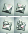
Four images of a BEV2 crystal during data collection. (a) Prior to any exposure. (b) After a 0.5 s exposure. (c) After a second 0.5 s exposure. (d) After a third 0.5 s exposure. The beam cross-section of 20 × 20 µm is shown.
Figure 4
Figure 4
A section of the 2.1 Å resolution 2F oF c electron-density map of BEV2 contoured at 1.5σ.
Figure 5
Figure 5
Comparison of in situ and frozen data collection from crystals of a protein–DNA complex. View of a crystal in a drop within a crystallization plate (a) and the diffraction obtained from this sample (b). Spots were observed to 10 Å resolution. A crystal grown in identical conditions was mounted in a fibre loop and cryocooled to 100 K (c). The resulting diffraction extended no further than 35 Å resolution (d) from the cryocooled crystal despite it being a larger size. In situ data collection allows a much more accurate assessment of crystallization conditions during the process of crystallization optimization.
Figure 6
Figure 6
On-axis microscope images of crystal hits and in situ diffraction patterns from three example membrane proteins. (a, d) A G-protein-coupled receptor protein crystallized in lipid cubic phase. (b, e) A complex of two domains of a bacterial membrane protein (the apparent misalignment of the beam centre and crystal in this case is a consequence of large refraction effects induced by the use of curved bottomed wells). (c, f) A bacterial membrane-anchor cytochrome protein. In (a), (b) and (c) the square box and cross-hair represent the beam size and position, respectively. In each case the size was 10 × 10 µm.
Figure 7
Figure 7
Strategy used for obtaining a data set from a large number of thin wedges (0.5–1.5°) of diffraction data.

References

    1. Adams, P. D. et al. (2010). Acta Cryst. D66, 213–221.
    1. Aishima, J., Owen, R. L., Axford, D., Shepherd, E., Winter, G., Levik, K., Gibbons, P., Ashton, A. & Evans, G. (2010). Acta Cryst. D66, 1032–1035. - PMC - PubMed
    1. Bingel-Erlenmeyer, R., Olieric, V., Grimshaw, J. P. A., Gabadinho, J., Wang, X., Ebner, S. G., Isenegger, A., Schneider, R., Schneider, J., Glettig, W., Pradervand, C., Panepucci, E. H., Tomizaki, T., Wang, M. & Schulze-Briese, C. (2011). Cryst. Growth Des. 11, 916–923.
    1. Brünger, A. T., Adams, P. D., Clore, G. M., DeLano, W. L., Gros, P., Grosse-Kunstleve, R. W., Jiang, J.-S., Kuszewski, J., Nilges, M., Pannu, N. S., Read, R. J., Rice, L. M., Simonson, T. & Warren, G. L. (1998). Acta Cryst. D54, 905–921. - PubMed
    1. Cockburn, J. J. B., Bamford, J. K. H., Grimes, J. M., Bamford, D. H. & Stuart, D. I. (2003). Acta Cryst. D59, 538–540. - PubMed

Publication types

MeSH terms