. 2012 Feb;38(1):13-27.
doi: 10.1016/j.rdc.2012.03.004. Epub 2012 Apr 12.

The vitamin D receptor: new paradigms for the regulation of gene expression by 1,25-dihydroxyvitamin D3

Affiliations

The vitamin D receptor: new paradigms for the regulation of gene expression by 1,25-dihydroxyvitamin D3

J Wesley Pike et al. Rheum Dis Clin North Am. 2012 Feb.

Abstract

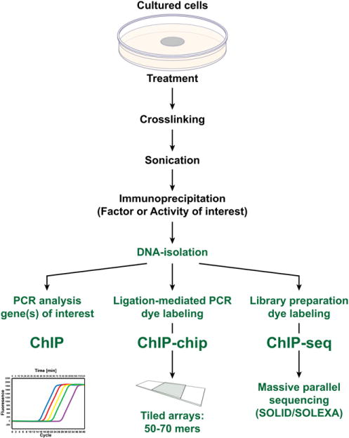
This article represents a summary of what is known of the VDR protein and its molecular mechanism of action at target genes. New methodologies now used, such as ChIP-chip and ChIP-seq, as well as novel reporter studies using large BAC clones stably transfected into culture cells or introduced as transgenes in mice, are providing new insights into how 1,25(OH)2D3-activated VDR modulates the expression of genes at single gene loci and at the level of gene networks. Many of these insights are unexpected and suggest that gene regulation is even more complex than previously appreciated. These studies also highlight new technologies and their central role in establishing fundamental biologic principles.


Figures

Fig. 1
Fig. 1
Structure and key features of the VDR. (A) The VDR protein comprised of a DNA-binding domain, a large ligand-binding domain, and a hinge region that links the 2 functional domains of the protein together. N, amino terminal end; C, carboxy terminal end; AF2, activation function 2. Amino acid numbers are shown. (B) Crystal structure of the VDR ligand-binding domain comprised of 12 α-helices (H1–H12). The N-terminal and C-terminal portions of the molecule are shown. A deletion in the molecular from G218 to M159 was required to achieve the formation of crystals. The position of 1,25(OH)2D3 is shown in the ligand-binding pocket as a stick figure. The ligand-binding domain was crystallized in the presence of a short peptide (indicated) representing a key LxxLL motif located in all coregulatory proteins that interact directly with the VDR. The repositioning of H12 as a consequence of 1,25(OH)2D3 binding provides the structural change necessary for interaction of the VDR with the LxxLL motif. (C) An electron density map of 1,25(OH)2D3 and adjacent amino acids within the VDR protein that make direct contact with the ligand. (Data from Vanhooke JL, Benning MM, Bauer CB, et al. Molecular structure of the rat vitamin D receptor ligand-binding domain complexed with 2-carbon-substituted vitamin D3 hormone analogues and a LXXLL-containing coactivator peptide. Biochemistry 2004;43(14):4101–10.)
Fig. 2
Fig. 2
Coregulatory complexes that are involved in mediating the actions of 1,25(OH)2D3 and the VDR. The general transcriptional apparatus is shown at the TSS and the VDR/RXR heterodimer is shown bound to its regulatory vitamin D response element or VDRE. Three regulatory complexes are shown that interact with the VDR: an ATPase-containing, chromatin remodeling complex termed SWI/SNF, a histone acetylation complex containing histone acetyltransferases (HAT) and Mediator complex. The latter facilitates the activation of RNA pol II through its C-terminal domain (CTD). Nucleosomes as well as individual proteins that comprise the individual coregulatory complexes are indicated.
Fig. 3
Fig. 3
Regulatory control of the synthesis (Cyp27b1), degradation (Cyp24a1), and mediation of activity (Vdr) of 1,25(OH)2D3 The concentration of 1,25(OH)2D3 in cells is determined through its synthesis and its degradation. Its functional activity is determined by the presence and intracellular concentration of the VDR.
Fig. 4
Fig. 4
Methodology associated with chromatin immunoprecipitation (ChIP) analysis and subsequent ChIP-DNA microarray (ChIP-chip) or massive parallel sequencing (ChIP-seq) analyses. Biologic samples are cross-linked, sonicated to prepare discrete size chromatin fragments, and then subjected to immunoprecipitation using selected antibodies. The precipitated DNA is then isolated and evaluated by polymerase chain reaction analysis or amplified and then subjected to either ChIP-chip or ChIP-seq analyses.

Republished from

References

    1. Haussler MR, Whitfield GK, Haussler CA, et al. The nuclear vitamin D receptor: biological and molecular regulatory properties revealed. J Bone Miner Res. 1998;13(3):325–49. - PubMed
    1. Sutton AL, MacDonald PN. Vitamin D: more than a “bone-a-fide” hormone. Mol Endocrinol. 2003;17(5):777–91. - PubMed
    1. Bouillon R, Carmeliet G, Verlinden L, et al. Vitamin D and human health: lessons from vitamin D receptor null mice. Endocr Rev. 2008;29(6):726–76. - PMC - PubMed
    1. Kim S, Yamazaki M, Zella LA, et al. Activation of receptor activator of NF-kappaB ligand gene expression by 1,25-dihydroxyvitamin D3 is mediated through multiple long-range enhancers. Mol Cell Biol. 2006;26(17):6469–86. - PMC - PubMed
    1. Zella LA, Kim S, Shevde NK, et al. Enhancers located within two introns of the vitamin D receptor gene mediate transcriptional autoregulation by 1,25-dihydrox-yvitamin D3. Mol Endocrinol. 2006;20(6):1231–47. - PubMed