. 2012 Jul;50(7):2524-30.
doi: 10.1016/j.fct.2012.04.010. Epub 2012 Apr 16.

Diallyl trisulfide as an inhibitor of benzo(a)pyrene-induced precancerous carcinogenesis in MCF-10A cells

Affiliations

Diallyl trisulfide as an inhibitor of benzo(a)pyrene-induced precancerous carcinogenesis in MCF-10A cells

Yasmeen M Nkrumah-Elie et al. Food Chem Toxicol. 2012 Jul.

Abstract

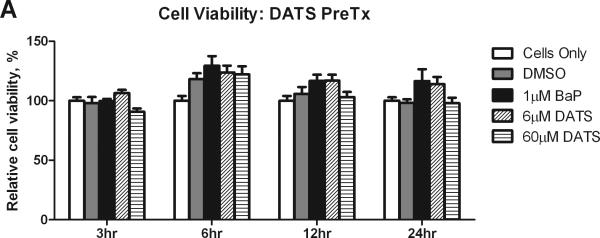
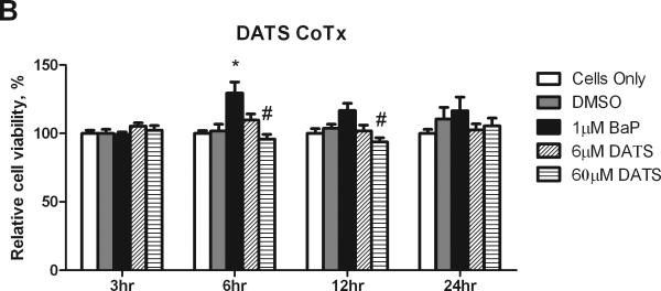
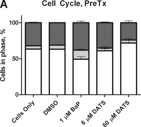
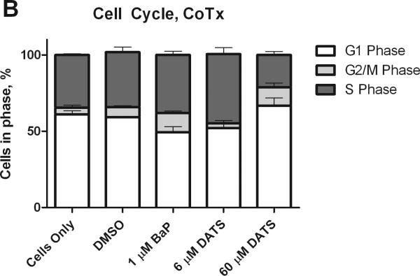
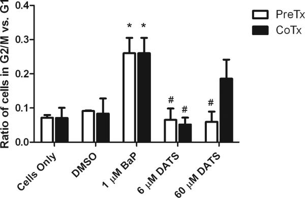
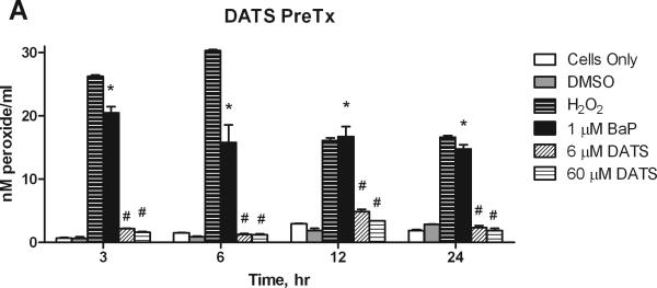
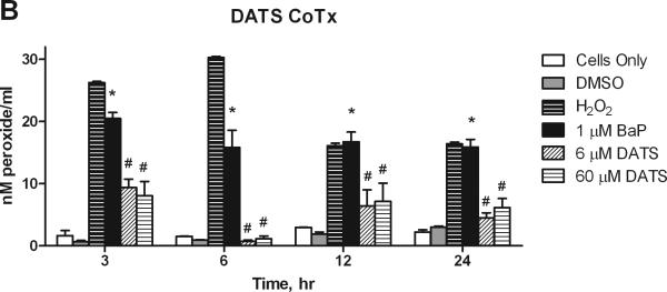
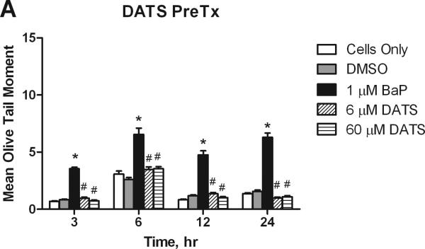
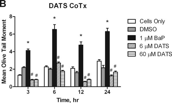
Diallyl trisulfide (DATS) is a garlic organosulfide that is toxic to cancer cells, however, little is known about its effect in the initiation phase of carcinogenesis. We sought to determine whether DATS could inhibit the carcinogen, benzo(a)pyrene (BaP), from inducing precancerous activity, in vitro. MCF-10A cells were either pre-treated (PreTx) or concurrently treated (CoTx) with 1 μM BaP, and 6 or 60 μM DATS for up to 24h. The DATS 6 and 60 μM CoTx inhibited BaP-induced cell proliferation by an average of 71.1% and 120.8%, respectively, at 6h. The 60 μM DATS pretreatment decreased BaP-induced G2/M cell cycle transition by 127%, and reduced the increase in cells in the S-phase by 42%; whereas 60 μM DATS CoTx induced a 177% increase in cells in G1. DATS effectively inhibited (P<0.001) BaP-induced peroxide formation by at least 54%, which may have prevented the formation of BaP-induced DNA strand breaks. In this study, we reveal mechanisms involved in DATS inhibition of BaP-induced carcinogenesis, including inhibition of cell proliferation, regulation of cell cycle, attenuation of ROS formation, and inhibition of DNA damage. At the doses evaluated, DATS appears to be an effective attenuator of BaP-induced breast carcinogenesis, in vitro.


Figures

Figure 1
Figure 1
Cell viability and proliferation resulting from Pre- and CoTx with DATS and BaP. MCF-10A cells were pretreated with DATS for four hours followed by the addition of 1 μM BaP (A) or treated concomitantly with 1 μM BaP and DATS (B). To determine cell viability, the MTS solution was applied to the cells for 2-3 hours and analyzed at an absorbance of 490nm. The absorbance of each treatment group was normalized relative to the cells only control for 100% cell viability. The graphs represent the average relative cell viability for eight replicates for N=3, +/- the SEM (* and # indicate a P<0.05 significant difference from the DMSO control and the 1 μM BaP only control, respectively).
Figure 1
Figure 1
Cell viability and proliferation resulting from Pre- and CoTx with DATS and BaP. MCF-10A cells were pretreated with DATS for four hours followed by the addition of 1 μM BaP (A) or treated concomitantly with 1 μM BaP and DATS (B). To determine cell viability, the MTS solution was applied to the cells for 2-3 hours and analyzed at an absorbance of 490nm. The absorbance of each treatment group was normalized relative to the cells only control for 100% cell viability. The graphs represent the average relative cell viability for eight replicates for N=3, +/- the SEM (* and # indicate a P<0.05 significant difference from the DMSO control and the 1 μM BaP only control, respectively).
Figure 2
Figure 2
Cell cycle analysis of MCF-10A cells treated with BaP and DATS. MCF-10A cells were either pretreated with DATS for four hours, followed by treatment with 1 μM BaP for 24 hours (A) or co-treated with DATS and BaP for 24 hours (B). The cells were fixed in ethanol, stained with propidium iodide, and analyzed by flow cytometry. The values represent the average percent of cells in each phase, G1, G2/M, and S, +/- SEM.
Figure 2
Figure 2
Cell cycle analysis of MCF-10A cells treated with BaP and DATS. MCF-10A cells were either pretreated with DATS for four hours, followed by treatment with 1 μM BaP for 24 hours (A) or co-treated with DATS and BaP for 24 hours (B). The cells were fixed in ethanol, stained with propidium iodide, and analyzed by flow cytometry. The values represent the average percent of cells in each phase, G1, G2/M, and S, +/- SEM.
Figure 3
Figure 3
The ratio of cells in G2/M and G1 phases. The bars indicate the average percentage of cells in G2/M divided by the average percentage of cells in G1, +/- SEM for the DATS PreTx and DATS and BaP CoTx after 24 hours. The * indicates a P<0.05 significant difference from the DMSO control, and # indicates a P<0.05 significant difference from the 1 μM BaP only control.
Figure 4
Figure 4
The suppression of BaP induced extracellular ROS formation by DATS. MCF-10A cells were exposed to a pretreatment of DATS followed by 1 μM BaP (A), or with a concomitant combination of 1 μM BaP and DATS (B), both for 3, 6, 12, or 24 hours. The results deem the average aqueous peroxides (as a model ROS), +/- SEM, as detected by the PeroxiDetect Kit in triplicate for N=3. The * indicates a P<0.05 significant difference from the DMSO control.
Figure 4
Figure 4
The suppression of BaP induced extracellular ROS formation by DATS. MCF-10A cells were exposed to a pretreatment of DATS followed by 1 μM BaP (A), or with a concomitant combination of 1 μM BaP and DATS (B), both for 3, 6, 12, or 24 hours. The results deem the average aqueous peroxides (as a model ROS), +/- SEM, as detected by the PeroxiDetect Kit in triplicate for N=3. The * indicates a P<0.05 significant difference from the DMSO control.
Figure 5
Figure 5
DATS inhibition of BaP induced DNA strand breaks. Utilizing the Comet assay, MCF-10A cells treated with a PreTx of DATS followed by BaP (A), or DATS and BaP concurrently (B) were analyzed for DNA single strand breaks and alkali liable sites. The results present the mean olive tail moment (OTM), as an indicator of DNA damage, +/- SEM for 150 cells for N=2. The * indicates a P<0.05 significant difference from the DMSO control.
Figure 5
Figure 5
DATS inhibition of BaP induced DNA strand breaks. Utilizing the Comet assay, MCF-10A cells treated with a PreTx of DATS followed by BaP (A), or DATS and BaP concurrently (B) were analyzed for DNA single strand breaks and alkali liable sites. The results present the mean olive tail moment (OTM), as an indicator of DNA damage, +/- SEM for 150 cells for N=2. The * indicates a P<0.05 significant difference from the DMSO control.

References

    1. Aboyade-Cole A, Darling-Reed S, Oriaku E, McCaskill M, Thomas R. Diallyl sulfide inhibits PhIP-induced cell death via the inhibition of DNA strand breaks in normal breast epithelial cells. Oncol Rep. 2008;20:319–323. - PMC - PubMed
    1. Ali M, Thompson M, Afzal M. Garlic and onions: their effect on eicosandoid metabolism and its clinical relevance. Prostaglandins Leukot Essent Fatty Acids. 2000;62:55–73. - PubMed
    1. Antosiewicz J, Herman-Antosiewicz A, Marynowski SW, Singh SV. 9C-Jun NH(2)-terminal kinase signaling axis regulates diallyl trisulfide-induced generation of reactive oxygen species and cell cycle arrest in human prostate cancer cells. Cancer Res. 2006;66:5379–5386. - PubMed
    1. Augusti KT, Mathew PT. Lipid lowering effect of allicin (diallyl disulfide oxide) on long-term feeding in normal rats. Experientia. 1974;30:468–470. - PubMed
    1. Block E. The chemistry of garlic and onions. Sci Am. 1985;252:114–119. - PubMed

Publication types

LinkOut - more resources