. 2012 Jun;123(6):861-72.
doi: 10.1007/s00401-012-0986-4. Epub 2012 Apr 22.

Neuromyelitis optica IgG and natural killer cells produce NMO lesions in mice without myelin loss

Affiliations

Neuromyelitis optica IgG and natural killer cells produce NMO lesions in mice without myelin loss

Julien Ratelade et al. Acta Neuropathol. 2012 Jun.

Abstract

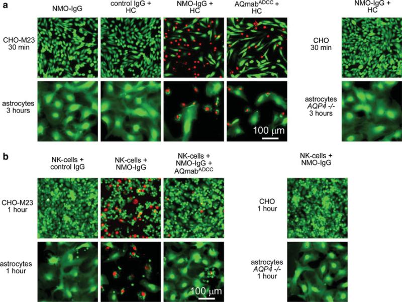
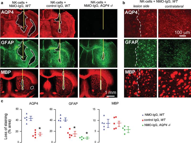
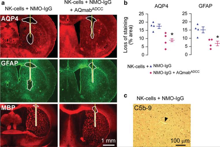
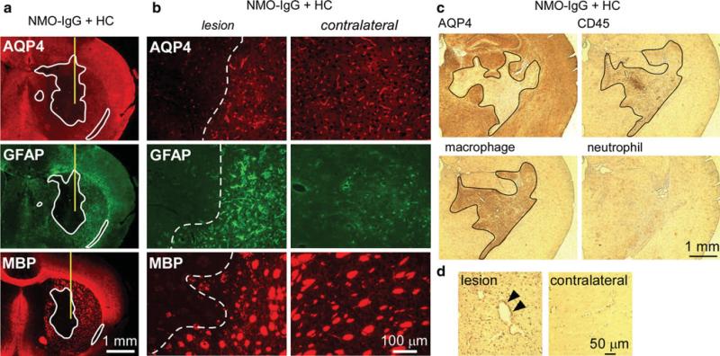
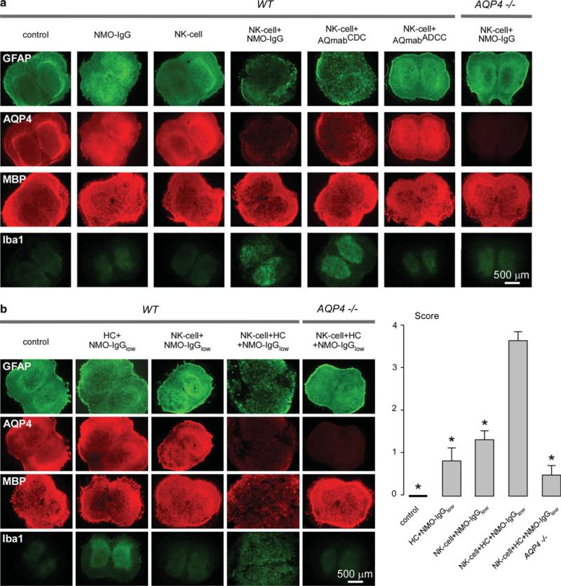
The pathogenesis of neuromyelitis optica (NMO) involves targeting of NMO-immunoglobulin G (NMO-IgG) to aquaporin-4 (AQP4) on astrocytes in the central nervous system. Prior work provided evidence for complement-dependent cytotoxicity (CDC) in NMO lesion development. Here, we show that antibody-dependent cellular cytotoxicity (ADCC), in the absence of complement, can also produce NMO-like lesions. Antibody-dependent cellular cytotoxicity was produced in vitro by incubation of mouse astrocyte cultures with human recombinant monoclonal NMO-IgG and human natural killer cells (NK-cells). Injection of NMO-IgG and NK-cells in mouse brain caused loss of AQP4 and GFAP, two characteristic features of NMO lesions, but little myelin loss. Lesions were minimal or absent following injection of: (1) control (non-NMO) IgG with NK-cells; (2) NMO-IgG and NK-cells in AQP4-deficient mice; or (3) NMO-IgG and NK-cells in wild-type mice together with an excess of mutated NMO-IgG lacking ADCC effector function. NK-cells greatly exacerbated NMO lesions produced by NMO-IgG and complement in an ex vivo spinal cord slice model of NMO, causing marked myelin loss. NMO-IgG can thus produce astrocyte injury by ADCC in a complement-independent and dependent manner, suggesting the potential involvement of ADCC in NMO pathogenesis.


Figures

Fig. 1
Fig. 1
NMO-IgG-dependent cytotoxicity in AQP4-transfected CHO cells and primary cultures of mouse astrocytes. a Fluorescence micrographs showing live/dead (green/red) staining of CHO cells expressing AQP4-M23 (CHO-M23) and mouse primary astrocyte cultures after incubation for indicated times with human complement (HC, 10 %) together with control (non-NMO) IgG or NMO-IgG or AQmabADCC (each 20 μg/mL). b Fluorescence micrographs showing live/dead staining of cells incubated with NK-cells (NK-cell:target cell ratio of 20:1) and control IgG or NMO-IgG (20 μg/mL) and/or AQmabADCC (200 μg/mL)
Fig. 2
Fig. 2
Intracerebral injection of NMO-IgG and NK-cells causes loss of AQP4 and GFAP but not of myelin. a Brains of wild type (WT) and AQP4 deficient (AQP4-/-) mice were injected with NK-cells (106 cells) and NMO-IgG or control IgG (4 μg). The mice were killed 4 days after injection and brain sections were immunostained for AQP4, GFAP and myelin (MBP). Yellow line represents the needle tract. White line shows the area of loss of immunoreactivity. Data are representative of four or five mice per group. b Higher magnification of WT mouse brain injected with NK-cells and NMO-IgG. Areas to the left of white dashed line show loss of AQP4 and GFAP but preservation of myelin. Contralateral (non-injected) hemispheres are shown (right). c Areas of loss of AQP4, GFAP and MBP immuno-reactivity (mean ± SE, *P < 0.01 compared with WT mice injected with NK-cells and NMO-IgG)
Fig. 3
Fig. 3
Intracerebral injection of NMO-IgG and NK-cells causes minimal inflammation. a Immunohistochemistry showing CD45-positive cell infiltration in the needle tract (black arrowheads) of WT mice injected with NMO-IgG or control IgG. Most infiltrating cells were positive for a macrophage marker but negative for a neutrophil marker. b Sections stained for microglia marker Iba1 showing microglial activation around the needle tract (labeled 1) in mice injected with NK-cells and NMO-IgG or control IgG compared to contralateral hemisphere (labeled 2). c Fluorescence micrographs of GFP-NK-cells 24 h and 4 days after intracerebral injection. White line represents the needle tract. Red arrowheads indicate infiltrating NK-cells in brain parenchyma. Micrographs at the bottom are a higher magnification of the sections at the top
Fig. 4
Fig. 4
Lesions following intracerebral injection of NMO-IgG and NK-cells are mediated by ADCC. a Brains of wild-type mice were injected with NK-cells (106 cells) and NMO-IgG (1 μg) or coinjected with NMO-IgG and an excess of AQmabADCC (5 μg). Yellow line represents the needle tract. White line shows the area of loss of immunoreactivity. Data are representative of four mice per group. b Quantification of areas of loss of AQP4 and GFAP immunoreactivity (mean ± SE, *P < 0.05 compared with mice injected with NK-cells and NMO-IgG). c C5b-9 staining of brain injected with NK-cells and NMO-IgG showing absence of complement activation. Arrowhead points to a brain vessel
Fig. 5
Fig. 5
Intracerebral injection of NMO-IgG and human complement produces demyelination and marked inflammation. a Brains were injected with NMO-IgG (0.4 μg) and human complement (3 μL) and stained for AQP4, GFAP and MBP. Yellow line represents the needle tract. White line shows areas of loss of immunoreactivity. Data are representative of four different mice. b Higher magnification of the sections shown in a. Areas to the left of white dashed lines show loss of AQP4, GFAP and myelin. Contralateral (non-injected) hemispheres are shown for comparison. c Same experiment as in (a) and (b), showing marked infiltration of CD45-positive cells in the area of AQP4 loss. Most of the infiltrating cells are positive for a macrophage marker but negative for a neutrophil marker. Black line outlines the lesion. d C5b-9 staining in lesion and in contralateral hemisphere. Arrowheads indicate perivascular complement deposition
Fig. 6
Fig. 6
NK-cells exacerbate NMO lesions produced by NMO-IgG and complement in an ex vivo spinal cord slice culture model. a Immunofluorescence of GFAP, AQP4, MBP and Iba1 in spinal cord slice cultures from wild type (WT) and AQP4-/- mice incubated with NMO-IgG, AQmabCDC or AQmabADCC (each 10 μg/mL) and/or NK-cells (106/well). Control indicates no NMO-IgG or NK-cells. b Same staining as in (a) of spinal cord slice cultures incubated with NK-cells (106/well) and/or human complement (HC, 5 %) and/or submaximal NMO-IgG (NMO-IgGlow, 3 μg/mL) (left). Scoring of NMO lesions (mean ± SE, 6–8 slices per condition, *P < 0.001 compared with NK-cell + HC + NMO-IgGlow, WT) (right)

References

    1. Alderson KL, Sondel PM. Clinical cancer therapy by NK cells via antibody-dependent cell-mediated cytotoxicity. J Biomed Biotechnol. 2011;2011:379123. - PMC - PubMed
    1. Becknell B, Caligiuri MA. Natural killer cells in innate immunity and cancer. J Immunother. 2008;31(8):685–692. - PubMed
    1. Bennett JL, Lam C, Kalluri SR, Saikali P, Bautista K, Dupree C, Glogowska M, Case D, Antel JP, Owens GP, Gilden D, Nessler S, Stadelmann C, Hemmer B. Intrathecal pathogenic anti-aquaporin-4 antibodies in early neuromyelitis optica. Ann Neurol. 2009;66(5):617–629. - PMC - PubMed
    1. Burgoon MP, Williamson RA, Owens GP, Ghausi O, Bastidas RB, Burton DR, Gilden DH. Cloning the antibody response in humans with inflammatory CNS disease: isolation of measles virus-specific antibodies from phage display libraries of a subacute sclerosing panencephalitis brain. J Neuroimmunol. 1999;94(1–2):204–211. - PubMed
    1. Capel PJ, van de Winkel JG, van den Herik-Oudijk IE, Verbeek JS. Heterogeneity of human IgG Fc receptors. Immunomethods. 1994;4(1):25–34. - PubMed

Publication types

MeSH terms

LinkOut - more resources