. 2012 Jun;403(6):1671-83.
doi: 10.1007/s00216-012-5968-9. Epub 2012 Apr 20.

Analysis of urinary oligosaccharides in lysosomal storage disorders by capillary high-performance anion-exchange chromatography-mass spectrometry

Affiliations

Analysis of urinary oligosaccharides in lysosomal storage disorders by capillary high-performance anion-exchange chromatography-mass spectrometry

Cees Bruggink et al. Anal Bioanal Chem. 2012 Jun.

Abstract

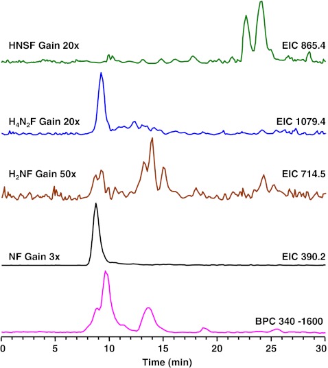
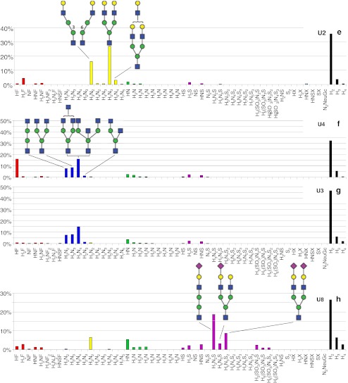
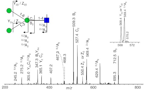
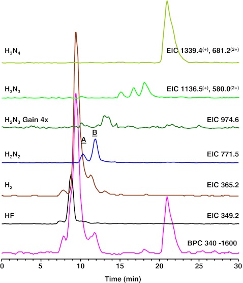
Many lysosomal storage diseases are characterized by an increased urinary excretion of glycoconjugates and oligosaccharides that are characteristic for the underlying enzymatic defect. Here, we have used capillary high-performance anion-exchange chromatography (HPAEC) hyphenated to mass spectrometry to analyze free oligosaccharides from urine samples of patients suffering from the lysosomal storage disorders fucosidosis, α-mannosidosis, G(M1)-gangliosidosis, G(M2)-gangliosidosis, and sialidosis. Glycan fingerprints were registered, and the patterns of accumulated oligosaccharides were found to reflect the specific blockages of the catabolic pathway. Our analytical approach allowed structural analysis of the excreted oligosaccharides and revealed several previously unpublished oligosaccharides. In conclusion, using online coupling of HPAEC with mass spectrometric detection, our study provides characteristic urinary oligosaccharide fingerprints with diagnostic potential for lysosomal storage disorders.


Figures

Fig. 1
Fig. 1
Separation of oligosaccharides in urine of a fucosidosis patient. H hexose, N N-acetylhexosamine, F fucose, S N-acetylneuraminic acid, BPC base peak chromatogram
Fig. 2
Fig. 2
Positive-ion fragmentation mass spectrum of the monosodiated disaccharide HexNAc1Fuc1 (precursor ion at m/z 390.2) from urine of a fucosidosis patient. Red triangle = fucose, blue square = N-acetylglucosamine
Fig. 3
Fig. 3
Histograms showing the relative abundance of the detected glycans in the urine sample (Table 1) of lysosomal storage disorders fucosidosis (a), α-mannosidosis (b, c, and d), GM1-gangliosidosis (e), GM2-gangliosidosis (f and g), and sialidosis (h). H or white circle = hexose, N or white square = N-acetylhexosamine, F or red triangle = fucose, S or purple diamond = N-acetylneuraminic acid, NeuGc = N-glycolylneuraminic acid, X = hexonic acid, SO 3 = sulfate, yellow circle = galactose, green circle = mannose, blue square = N-acetylglucosamine
Fig. 3
Fig. 3
Histograms showing the relative abundance of the detected glycans in the urine sample (Table 1) of lysosomal storage disorders fucosidosis (a), α-mannosidosis (b, c, and d), GM1-gangliosidosis (e), GM2-gangliosidosis (f and g), and sialidosis (h). H or white circle = hexose, N or white square = N-acetylhexosamine, F or red triangle = fucose, S or purple diamond = N-acetylneuraminic acid, NeuGc = N-glycolylneuraminic acid, X = hexonic acid, SO 3 = sulfate, yellow circle = galactose, green circle = mannose, blue square = N-acetylglucosamine
Fig. 4
Fig. 4
Positive-ion fragmentation mass spectrum of the monosodiated tetrasaccharide Man3GlcNAc1 (precursor ion at m/z 730.6) from urine of a α-mannosidosis patient. Green circle = mannose, blue square = N-acetylglucosamine
Fig. 5
Fig. 5
Separation of oligosaccharides in urine of a GM1-gangliosidosis patient. H hexose, N N-acetylhexosamine, BPC base peak chromatogram
Fig. 6
Fig. 6
Positive-ion fragmentation mass spectrum of the disodiated diantennary oligosaccharide Hex5HexNAc3 (precursor ion at m/z 742.1) from urine of a GM1-gangliosidosis patient. Yellow circle galactose, green circle mannose, blue square N-acetylglucosamine
Fig. 7
Fig. 7
Separation of oligosaccharides in urine of a GM2-gangliosisis patient. Fragment ion spectra of the species A and B is shown in Fig. 8. F fucose, H hexose, N N-acetylhexosamine, BPC base peak chromatogram
Fig. 8
Fig. 8
Positive-ion fragmentation mass spectra of two isomeric monosodiated tetrasaccharides Hex2HexNAc2 (precursor ion at m/z 771.5) from urine of a GM2-gangliosisis patient. The separation of A and B is shown in Fig. 7. Green circle mannose, blue square = N-acetylglucosamine

References

    1. Cantz M, Ulrich-Bott B. Disorders of glycoprotein degradation. J Inherit Metab Dis. 1990;13:523–537. doi: 10.1007/BF01799510. - DOI - PubMed
    1. Lundblad A, Lundsten J, Nordén NE, Sjöblad S, Svensson S, Öckerman PA, Gehlhoff M. Urinary abnormalities in fucosidosis. Eur J Biochem. 1978;83:513–521. doi: 10.1111/j.1432-1033.1978.tb12118.x. - DOI - PubMed
    1. Ockerman PA. Diseases of glycoprotein storage. Lancet. 1969;1:734. doi: 10.1016/S0140-6736(69)92687-7. - DOI - PubMed
    1. Ockerman PA, Autio S, Norden NE. Diagnosis of mannosidosis. Lancet. 1973;1:207–208. doi: 10.1016/S0140-6736(73)90045-7. - DOI - PubMed
    1. Winchester B. Lysosomal metabolism of glycoproteins. Glycobiology. 2005;15:1R–15R. doi: 10.1093/glycob/cwi041. - DOI - PubMed

MeSH terms

LinkOut - more resources