. 2012 Jul 18;18(1):851-60.
doi: 10.2119/molmed.2012.00011.

Mammalian DNA is an endogenous danger signal that stimulates local synthesis and release of complement factor B

Affiliations

Mammalian DNA is an endogenous danger signal that stimulates local synthesis and release of complement factor B

David J Kaczorowski et al. Mol Med. .

Abstract

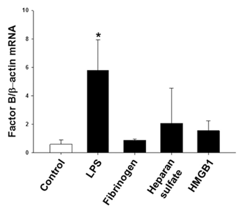
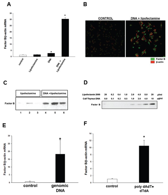
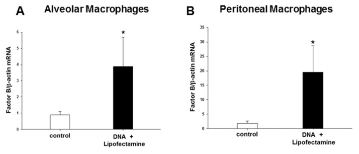
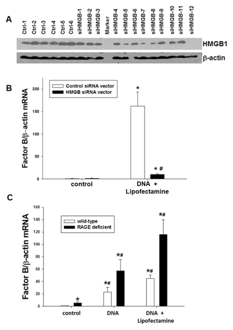
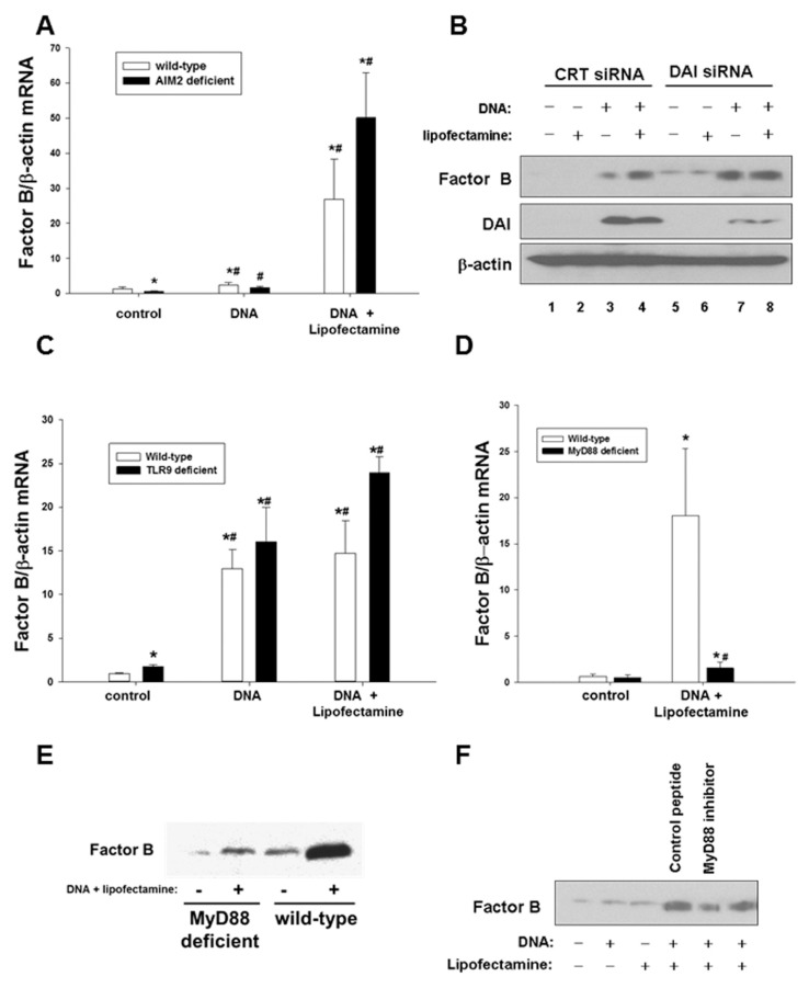
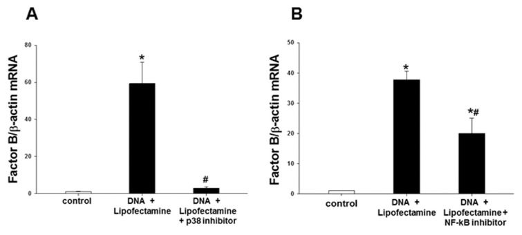
Complement factor B plays a critical role in ischemic tissue injury and autoimmunity. Factor B is dynamically synthesized and released by cells outside of the liver, but the molecules that trigger local factor B synthesis and release during endogenous tissue injury have not been identified. We determined that factor B is upregulated early after cold ischemia-reperfusion in mice, using a heterotopic heart transplant model. These data suggested upregulation of factor B by damage-associated molecular patterns (DAMPs), but multiple common DAMPs did not induce factor B in RAW264.7 mouse macrophages. However, exogenous DNA induced factor B mRNA and protein expression in RAW cells in vitro, as well as in peritoneal and alveolar macrophages in vivo. To determine the cellular mechanisms involved in DNA-induced factor B upregulation we then investigated the role of multiple known DNA receptors or binding partners. We stimulated peritoneal macrophages from wild-type (WT), toll-like receptor 9 (TLR9)-deficient, receptor for advanced glycation end products (RAGE)⁻/⁻ and myeloid differentiation factor 88 (MyD88)⁻/⁻ mice, or mouse macrophages deficient in high-mobility group box proteins (HMGBs), DNA-dependent activator of interferon-regulatory factors (DAI) or absent in melanoma 2 (AIM2), with DNA in the presence or absence of lipofection reagent. Reverse transcription-polymerase chain reaction, Western blotting and immunocytochemical analysis were employed for analysis. Synthesis of factor B was independent of TLR9, RAGE, DAI and AIM2, but was dependent on HMGBs, MyD88, p38 and NF-κB. Our data therefore show that mammalian DNA is an endogenous molecule that stimulates factor B synthesis and release from macrophages via HMGBs, MyD88, p38 and NF-κB signaling. This activation of the immune system likely contributes to damage following sterile injury such as hemorrhagic shock and ischemia-reperfusion.


Figures

Figure 1
Figure 1
Factor B is not upregulated in macrophages by common known TLR4-mediated DAMPs. Expression of factor B mRNA in RAW264.7 macrophages treated with LPS (positive control), or DAMPs (fibrinogen, heparan sulfate, HMGB1) or PBS (control) for 6 h. *p < 0.05 versus control. Experiment performed in triplicate and repeated at least three times.
Figure 2
Figure 2
DNA stimulates factor B synthesis and release by macrophages in vitro. (A) Factor B mRNA expression in RAW264.7 macrophages treated for 6 h with Lipofectamine 2000 alone, calf thymus DNA (10 μg/mL) alone or calf thymus DNA (10 μg/mL) complexed with Lipofectamine 2000. *p < 0.001 versus control. (B) Immunohistochemistry for factor B and β-actin in RAW264.7 macrophages treated with DNA (10 μg/mL) complexed with Lipofectamine 2000, or Lipofectamine 2000 alone (control) for 6 h. (C) Factor B release into supernatants from RAW264.7 macrophages treated with DNA (10 μg/mL) complexed with Lipofectamine 2000 or Lipofectamine 2000 alone for 24 h. (D) Factor B protein expression in cell culture supernatants from RAW264.7 macrophages treated with DNA complexed with Lipofectamine 2000, or Lipofectamine 2000 alone for 24 h at various concentrations as indicated. (E) Expression of factor B mRNA in RAW264.7 macrophages treated for 6 h with mouse genomic DNA (10 μg/mL) complexed with Lipofectamine 2000. *p < 0.001 versus control. (F) Factor B mRNA expression in RAW264.7 macrophages treated for 6 h with poly (dAdT)·poly(dTdA) (10 μg/mL) complexed with lipofection reagent. *p < 0.001 versus control. All experiments performed in triplicate, and repeated at least three times.
Figure 3
Figure 3
DNA stimulates factor B upregulation in vivo. (A) Factor B mRNA expression in alveolar macrophages harvested 6 h after intratracheal injection of either Lipofectamine 2000 in carrier or calf thymus DNA complexed with Lipofectamine 2000. *p < 0.005 versus control. (B) Factor B mRNA expression in peritoneal macrophages harvested at 6 h after intraperitoneal injections of either Lipofectamine 2000 in carrier, or calf thymus DNA complexed with Lipofectamine 2000. *p < 0.001 versus control; n = 6 per experimental group and experiments were repeated at least twice.
Figure 4
Figure 4
Factor B synthesis and release after stimulation with DNA requires HMGBs but not RAGE A. Expression of HMGB1 in RAW264.7 cell clones stably transfected with siRNA against HMGBs (siHMGB), or control siRNA (Ctrl). (A) Clonal cell line (siHMGB-12) expressing relatively low levels of HMGB1 was selected for future experiments. (B.) Factor B mRNA expression in control or siHMGB-12 cells 6 h after stimulation with calf thymus DNA (10 μg/mL) complexed with Lipofectamine 2000. *p < 0.001 versus unstimulated cells containing control siRNA vector, #p < 0.001 versus cells containing control siRNA vector stimulated with DNA/lipofectamine. (C) Factor B mRNA expression in peritoneal macrophages from WT and RAGE-deficient mice after stimulation with calf thymus DNA (10 μg/mL) alone, or calf thymus DNA (10 μg/mL) complexed with Lipofectamine 2000 for 6 h. *p < 0.01 versus WT control, #p < 0.01 versus unstimulated RAGE-deficient cells. Experiments were performed in triplicate and repeated at least three times.
Figure 5
Figure 5
Factor B upregulation in macrophages after stimulation with DNA is independent of AIM2, DAI and TLR9, but dependent on MyD88. (A) Factor B mRNA expression in WT and AIM2-deficient mouse peritoneal macrophages 6 h after stimulation with calf thymus DNA (10 μg/mL) alone or calf thymus DNA (10 μg/mL) complexed with Lipofectamine 2000. *p < 0.001 versus WT control, #p < 0.001 versus unstimulated AIM2-deficient cells. (B) Expression of factor B and DAI in cell lysates of RAW264.7 macrophages pre-treated with either control (CRT) or DAI-targeting siRNA and then stimulated for 24 h with DNA alone (10 μg/mL), Lipofectamine 2000 alone or DNA (10 μg/mL) complexed with Lipofectatmine 2000 as indicated. (C) Expression of factor B mRNA in peritoneal macrophages from WT and TLR9-deficient mice stimulated with calf thymus DNA (10 μg/mL) alone or calf thymus DNA (10 μg/mL) complexed with Lipofectamine 2000 for 6 h. *p < 0.001 versus WT control, #p < 0.001 versus unstimulated TLR9-deficient cells. (D) Factor B mRNA expression in peritoneal macrophages from WT and MyD88-deficient mice 6 h after stimulation with calf thymus DNA (10 μg/mL) complexed with Lipofectamine 2000. *p < 0.001 versus WT control, #p < 0.001 versus stimulated WT cells. (E) Factor B release into cell culture supernatants of peritoneal macrophages from WT and MyD88-deficient mice stimulated with calf thymus DNA (10 μg/mL) complexed with Lipofectamine 2000 for 6 h. (F) Factor B mRNA expression in cell culture supernatants from RAW264.7 macrophages pretreated with MyD88-inhibitor peptide or control peptide, then stimulated with DNA alone (10 μg/mL), Lipofectamine 2000 alone or with DNA (10 μg/mL) complexed with Lipofectatmine 2000 for 6 h as indicated. Experiments were performed in triplicate and repeated at least three times.
Figure 6
Figure 6
p38 and NF-κB mediate factor B production in macrophages. (A) Expression of factor B mRNA in RAW264.7 macrophages pretreated for 30 min with p38 inhibitor (SB 203580, 40 μmol/L) then treated with DNA (10 μg/mL) complexed with Lipofectamine 2000 for 6h. *p < 0.001 versus control, #p < 0.001 versus DNA + Lipofectamine 2000. (B) Factor B mRNA expression in RAW264.7 macrophages pretreated with NF-κB activation inhibitor (10 μmol/L) overnight, then treated with DNA (10 μg/mL) complexed with Lipofectamine 2000 for 6 h. *p < 0.001 versus control, #p < 0.001 versus DNA + Lipofectamine 2000. Experiments were performed in triplicate and repeated at least three times.

References

    1. Arumugam TV, Shiels IA, Woodruff TM, Granger DN, Taylor SM. The role of the complement system in ischemia-reperfusion injury. Shock. 2004;21:401–9. - PubMed
    1. Diepenhorst GM, van Gulik TM, Hack CE. Complement-mediated ischemia-reperfusion injury: lessons learned from animal and clinical studies. Ann Surg. 2009;249:889–99. - PubMed
    1. Brooimans RA, et al. Interleukin 2 mediates stimulation of complement C3 biosynthesis in human proximal tubular epithelial cells. J Clin Invest. 1991;88:379–84. - PMC - PubMed
    1. Kawakami Y, et al. TNF-alpha stimulates the biosynthesis of complement C3 and factor B by human umbilical cord vein endothelial cells. Cancer Lett. 1997;116:21–6. - PubMed
    1. McPhaden AR, Whaley K. Complement biosynthesis by mononuclear phagocytes. Immunol Res. 1993;12:213–32. - PubMed

Publication types

MeSH terms

LinkOut - more resources